Iodine »
PDB 1a31-1ga5 »
1c3x »
Iodine in PDB 1c3x: Purine Nucleoside Phosphorylase From Cellulomonas Sp. in Complex with 8-Iodo-Guanine
Protein crystallography data
The structure of Purine Nucleoside Phosphorylase From Cellulomonas Sp. in Complex with 8-Iodo-Guanine, PDB code: 1c3x
was solved by
J.Tebbe,
A.Bzowska,
B.Wielgus-Kutrowska,
W.Schroeder,
Z.Kazimierczuk,
D.Shugar,
W.Saenger,
G.Koellner,
with X-Ray Crystallography technique. A brief refinement statistics is given in the table below:
| Resolution Low / High (Å) | 44.00 / 2.40 |
| Space group | P 21 21 21 |
| Cell size a, b, c (Å), α, β, γ (°) | 63.319, 108.340, 117.562, 90.00, 90.00, 90.00 |
| R / Rfree (%) | 20 / 25.1 |
Other elements in 1c3x:
The structure of Purine Nucleoside Phosphorylase From Cellulomonas Sp. in Complex with 8-Iodo-Guanine also contains other interesting chemical elements:
| Calcium | (Ca) | 1 atom |
Iodine Binding Sites:
The binding sites of Iodine atom in the Purine Nucleoside Phosphorylase From Cellulomonas Sp. in Complex with 8-Iodo-Guanine
(pdb code 1c3x). This binding sites where shown within
5.0 Angstroms radius around Iodine atom.
In total 3 binding sites of Iodine where determined in the Purine Nucleoside Phosphorylase From Cellulomonas Sp. in Complex with 8-Iodo-Guanine, PDB code: 1c3x:
Jump to Iodine binding site number: 1; 2; 3;
In total 3 binding sites of Iodine where determined in the Purine Nucleoside Phosphorylase From Cellulomonas Sp. in Complex with 8-Iodo-Guanine, PDB code: 1c3x:
Jump to Iodine binding site number: 1; 2; 3;
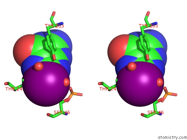
Iodine binding site 1 out of 3 in 1c3x
Go back to
Iodine binding site 1 out
of 3 in the Purine Nucleoside Phosphorylase From Cellulomonas Sp. in Complex with 8-Iodo-Guanine
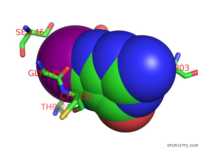
Mono view

Stereo pair view
Mono view
Stereo pair view
A full contact list of Iodine with other atoms in the I binding
site number 1 of Purine Nucleoside Phosphorylase From Cellulomonas Sp. in Complex with 8-Iodo-Guanine within 5.0Å range:
|
Iodine binding site 2 out of 3 in 1c3x
Go back to
Iodine binding site 2 out
of 3 in the Purine Nucleoside Phosphorylase From Cellulomonas Sp. in Complex with 8-Iodo-Guanine

Mono view
Stereo pair view
Mono view
Stereo pair view
A full contact list of Iodine with other atoms in the I binding
site number 2 of Purine Nucleoside Phosphorylase From Cellulomonas Sp. in Complex with 8-Iodo-Guanine within 5.0Å range:
|
Iodine binding site 3 out of 3 in 1c3x
Go back to
Iodine binding site 3 out
of 3 in the Purine Nucleoside Phosphorylase From Cellulomonas Sp. in Complex with 8-Iodo-Guanine

Mono view

Stereo pair view
Mono view
Stereo pair view
A full contact list of Iodine with other atoms in the I binding
site number 3 of Purine Nucleoside Phosphorylase From Cellulomonas Sp. in Complex with 8-Iodo-Guanine within 5.0Å range:
|
Reference:
J.Tebbe,
A.Bzowska,
B.Wielgus-Kutrowska,
W.Schroder,
Z.Kazimierczuk,
D.Shugar,
W.Saenger,
G.Koellner.
Crystal Structure of the Purine Nucleoside Phosphorylase (Pnp) From Cellulomonas Sp. and Its Implication For the Mechanism of Trimeric Pnps. J.Mol.Biol. V. 294 1239 1999.
ISSN: ISSN 0022-2836
PubMed: 10600382
DOI: 10.1006/JMBI.1999.3327
Page generated: Sun Aug 11 09:25:58 2024
ISSN: ISSN 0022-2836
PubMed: 10600382
DOI: 10.1006/JMBI.1999.3327
Last articles
Zn in 9JYWZn in 9IR4
Zn in 9IR3
Zn in 9GMX
Zn in 9GMW
Zn in 9JEJ
Zn in 9ERF
Zn in 9ERE
Zn in 9EGV
Zn in 9EGW