Iodine »
PDB 1a31-1ga5 »
1cel »
Iodine in PDB 1cel: The Three-Dimensional Crystal Structure of the Catalytic Core of Cellobiohydrolase I From Trichoderma Reesei
Enzymatic activity of The Three-Dimensional Crystal Structure of the Catalytic Core of Cellobiohydrolase I From Trichoderma Reesei
All present enzymatic activity of The Three-Dimensional Crystal Structure of the Catalytic Core of Cellobiohydrolase I From Trichoderma Reesei:
3.2.1.91;
3.2.1.91;
Protein crystallography data
The structure of The Three-Dimensional Crystal Structure of the Catalytic Core of Cellobiohydrolase I From Trichoderma Reesei, PDB code: 1cel
was solved by
C.Divne,
T.A.Jones,
with X-Ray Crystallography technique. A brief refinement statistics is given in the table below:
| Resolution Low / High (Å) | 7.50 / 1.80 |
| Space group | P 21 21 2 |
| Cell size a, b, c (Å), α, β, γ (°) | 84.000, 86.200, 111.800, 90.00, 90.00, 90.00 |
| R / Rfree (%) | 18.1 / n/a |
Other elements in 1cel:
The structure of The Three-Dimensional Crystal Structure of the Catalytic Core of Cellobiohydrolase I From Trichoderma Reesei also contains other interesting chemical elements:
| Calcium | (Ca) | 1 atom |
Iodine Binding Sites:
The binding sites of Iodine atom in the The Three-Dimensional Crystal Structure of the Catalytic Core of Cellobiohydrolase I From Trichoderma Reesei
(pdb code 1cel). This binding sites where shown within
5.0 Angstroms radius around Iodine atom.
In total 2 binding sites of Iodine where determined in the The Three-Dimensional Crystal Structure of the Catalytic Core of Cellobiohydrolase I From Trichoderma Reesei, PDB code: 1cel:
Jump to Iodine binding site number: 1; 2;
In total 2 binding sites of Iodine where determined in the The Three-Dimensional Crystal Structure of the Catalytic Core of Cellobiohydrolase I From Trichoderma Reesei, PDB code: 1cel:
Jump to Iodine binding site number: 1; 2;
Iodine binding site 1 out of 2 in 1cel
Go back to
Iodine binding site 1 out
of 2 in the The Three-Dimensional Crystal Structure of the Catalytic Core of Cellobiohydrolase I From Trichoderma Reesei
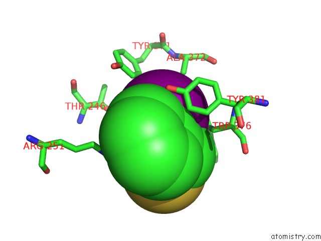
Mono view
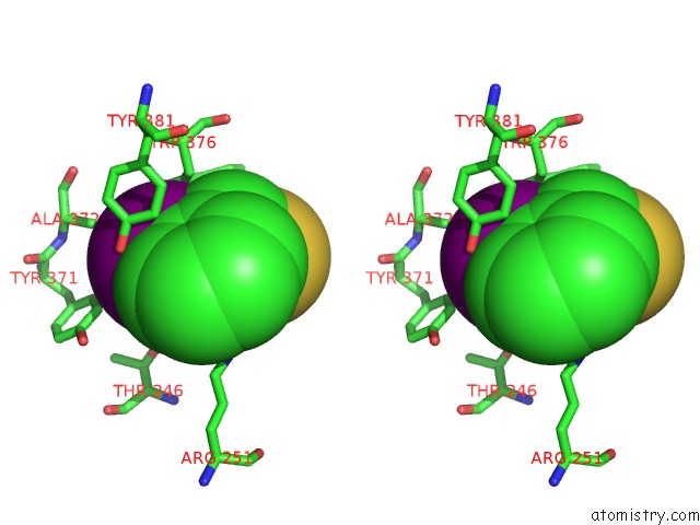
Stereo pair view
Mono view
Stereo pair view
A full contact list of Iodine with other atoms in the I binding
site number 1 of The Three-Dimensional Crystal Structure of the Catalytic Core of Cellobiohydrolase I From Trichoderma Reesei within 5.0Å range:
|
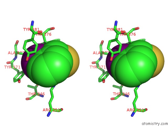
Iodine binding site 2 out of 2 in 1cel
Go back to
Iodine binding site 2 out
of 2 in the The Three-Dimensional Crystal Structure of the Catalytic Core of Cellobiohydrolase I From Trichoderma Reesei
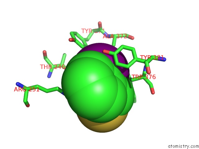
Mono view
Stereo pair view
Mono view
Stereo pair view
A full contact list of Iodine with other atoms in the I binding
site number 2 of The Three-Dimensional Crystal Structure of the Catalytic Core of Cellobiohydrolase I From Trichoderma Reesei within 5.0Å range:
|
Reference:
C.Divne,
J.Stahlberg,
T.Reinikainen,
L.Ruohonen,
G.Pettersson,
J.K.Knowles,
T.T.Teeri,
T.A.Jones.
The Three-Dimensional Crystal Structure of the Catalytic Core of Cellobiohydrolase I From Trichoderma Reesei. Science V. 265 524 1994.
ISSN: ISSN 0036-8075
PubMed: 8036495
Page generated: Sun Aug 11 09:29:53 2024
ISSN: ISSN 0036-8075
PubMed: 8036495
Last articles
Zn in 9J0NZn in 9J0O
Zn in 9J0P
Zn in 9FJX
Zn in 9EKB
Zn in 9C0F
Zn in 9CAH
Zn in 9CH0
Zn in 9CH3
Zn in 9CH1