Iodine »
PDB 1a31-1ga5 »
1etb »
Iodine in PDB 1etb: The X-Ray Crystal Structure Refinements of Normal Human Transthyretin and the Amyloidogenic Val 30-->Met Variant to 1.7 Angstroms Resolution
Protein crystallography data
The structure of The X-Ray Crystal Structure Refinements of Normal Human Transthyretin and the Amyloidogenic Val 30-->Met Variant to 1.7 Angstroms Resolution, PDB code: 1etb
was solved by
B.C.Braden,
L.K.Steinrauf,
J.A.Hamilton,
with X-Ray Crystallography technique. A brief refinement statistics is given in the table below:
| Resolution Low / High (Å) | 5.00 / 1.70 |
| Space group | P 21 21 2 |
| Cell size a, b, c (Å), α, β, γ (°) | 43.720, 86.090, 65.790, 90.00, 90.00, 90.00 |
| R / Rfree (%) | n/a / n/a |
Iodine Binding Sites:
The binding sites of Iodine atom in the The X-Ray Crystal Structure Refinements of Normal Human Transthyretin and the Amyloidogenic Val 30-->Met Variant to 1.7 Angstroms Resolution
(pdb code 1etb). This binding sites where shown within
5.0 Angstroms radius around Iodine atom.
In total 8 binding sites of Iodine where determined in the The X-Ray Crystal Structure Refinements of Normal Human Transthyretin and the Amyloidogenic Val 30-->Met Variant to 1.7 Angstroms Resolution, PDB code: 1etb:
Jump to Iodine binding site number: 1; 2; 3; 4; 5; 6; 7; 8;
In total 8 binding sites of Iodine where determined in the The X-Ray Crystal Structure Refinements of Normal Human Transthyretin and the Amyloidogenic Val 30-->Met Variant to 1.7 Angstroms Resolution, PDB code: 1etb:
Jump to Iodine binding site number: 1; 2; 3; 4; 5; 6; 7; 8;
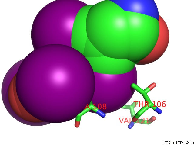
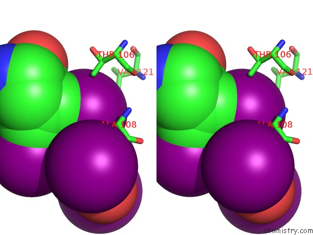
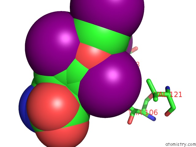
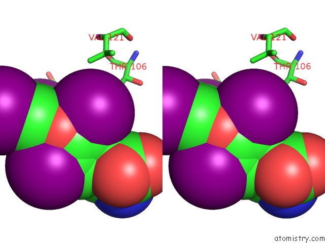
Iodine binding site 1 out of 8 in 1etb
Go back to
Iodine binding site 1 out
of 8 in the The X-Ray Crystal Structure Refinements of Normal Human Transthyretin and the Amyloidogenic Val 30-->Met Variant to 1.7 Angstroms Resolution
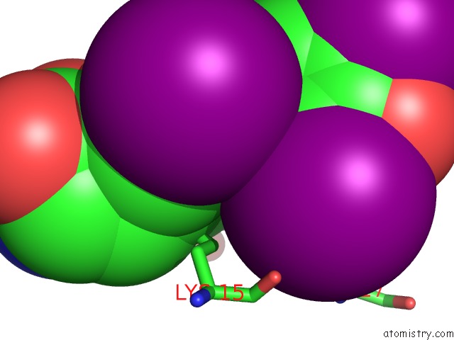
Mono view
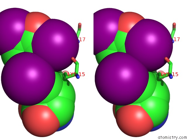
Stereo pair view
Mono view
Stereo pair view
A full contact list of Iodine with other atoms in the I binding
site number 1 of The X-Ray Crystal Structure Refinements of Normal Human Transthyretin and the Amyloidogenic Val 30-->Met Variant to 1.7 Angstroms Resolution within 5.0Å range:
|
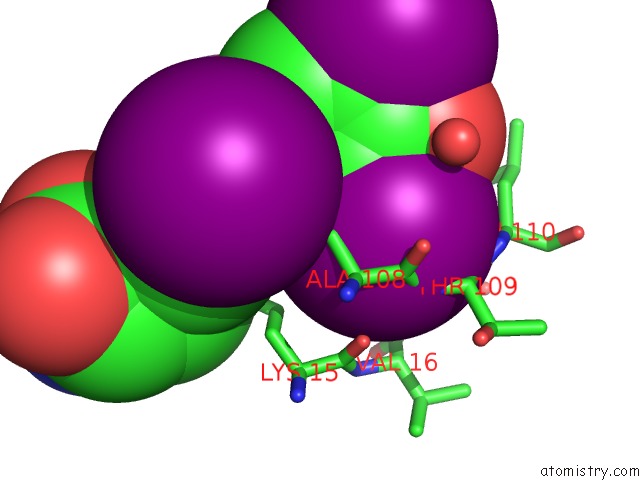
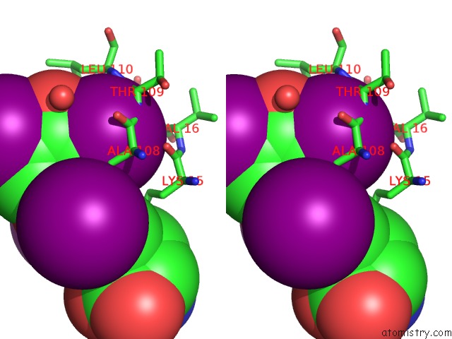
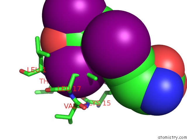
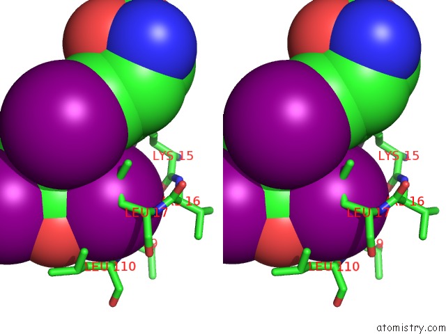
Iodine binding site 2 out of 8 in 1etb
Go back to
Iodine binding site 2 out
of 8 in the The X-Ray Crystal Structure Refinements of Normal Human Transthyretin and the Amyloidogenic Val 30-->Met Variant to 1.7 Angstroms Resolution
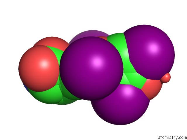
Mono view
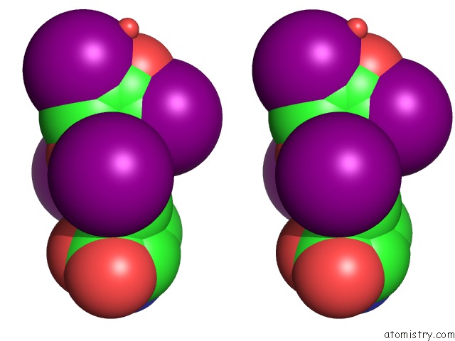
Stereo pair view
Mono view
Stereo pair view
A full contact list of Iodine with other atoms in the I binding
site number 2 of The X-Ray Crystal Structure Refinements of Normal Human Transthyretin and the Amyloidogenic Val 30-->Met Variant to 1.7 Angstroms Resolution within 5.0Å range:
|
Iodine binding site 3 out of 8 in 1etb
Go back to
Iodine binding site 3 out
of 8 in the The X-Ray Crystal Structure Refinements of Normal Human Transthyretin and the Amyloidogenic Val 30-->Met Variant to 1.7 Angstroms Resolution
Mono view
Stereo pair view
Mono view
Stereo pair view
A full contact list of Iodine with other atoms in the I binding
site number 3 of The X-Ray Crystal Structure Refinements of Normal Human Transthyretin and the Amyloidogenic Val 30-->Met Variant to 1.7 Angstroms Resolution within 5.0Å range:
|
Iodine binding site 4 out of 8 in 1etb
Go back to
Iodine binding site 4 out
of 8 in the The X-Ray Crystal Structure Refinements of Normal Human Transthyretin and the Amyloidogenic Val 30-->Met Variant to 1.7 Angstroms Resolution
Mono view
Stereo pair view
Mono view
Stereo pair view
A full contact list of Iodine with other atoms in the I binding
site number 4 of The X-Ray Crystal Structure Refinements of Normal Human Transthyretin and the Amyloidogenic Val 30-->Met Variant to 1.7 Angstroms Resolution within 5.0Å range:
|
Iodine binding site 5 out of 8 in 1etb
Go back to
Iodine binding site 5 out
of 8 in the The X-Ray Crystal Structure Refinements of Normal Human Transthyretin and the Amyloidogenic Val 30-->Met Variant to 1.7 Angstroms Resolution
Mono view
Stereo pair view
Mono view
Stereo pair view
A full contact list of Iodine with other atoms in the I binding
site number 5 of The X-Ray Crystal Structure Refinements of Normal Human Transthyretin and the Amyloidogenic Val 30-->Met Variant to 1.7 Angstroms Resolution within 5.0Å range:
|
Iodine binding site 6 out of 8 in 1etb
Go back to
Iodine binding site 6 out
of 8 in the The X-Ray Crystal Structure Refinements of Normal Human Transthyretin and the Amyloidogenic Val 30-->Met Variant to 1.7 Angstroms Resolution
Mono view
Stereo pair view
Mono view
Stereo pair view
A full contact list of Iodine with other atoms in the I binding
site number 6 of The X-Ray Crystal Structure Refinements of Normal Human Transthyretin and the Amyloidogenic Val 30-->Met Variant to 1.7 Angstroms Resolution within 5.0Å range:
|
Iodine binding site 7 out of 8 in 1etb
Go back to
Iodine binding site 7 out
of 8 in the The X-Ray Crystal Structure Refinements of Normal Human Transthyretin and the Amyloidogenic Val 30-->Met Variant to 1.7 Angstroms Resolution
Mono view
Stereo pair view
Mono view
Stereo pair view
A full contact list of Iodine with other atoms in the I binding
site number 7 of The X-Ray Crystal Structure Refinements of Normal Human Transthyretin and the Amyloidogenic Val 30-->Met Variant to 1.7 Angstroms Resolution within 5.0Å range:
|
Iodine binding site 8 out of 8 in 1etb
Go back to
Iodine binding site 8 out
of 8 in the The X-Ray Crystal Structure Refinements of Normal Human Transthyretin and the Amyloidogenic Val 30-->Met Variant to 1.7 Angstroms Resolution
Mono view
Stereo pair view
Mono view
Stereo pair view
A full contact list of Iodine with other atoms in the I binding
site number 8 of The X-Ray Crystal Structure Refinements of Normal Human Transthyretin and the Amyloidogenic Val 30-->Met Variant to 1.7 Angstroms Resolution within 5.0Å range:
|
Reference:
J.A.Hamilton,
L.K.Steinrauf,
B.C.Braden,
J.Liepnieks,
M.D.Benson,
G.Holmgren,
O.Sandgren,
L.Steen.
The X-Ray Crystal Structure Refinements of Normal Human Transthyretin and the Amyloidogenic Val-30-->Met Variant to 1.7-A Resolution. J.Biol.Chem. V. 268 2416 1993.
ISSN: ISSN 0021-9258
PubMed: 8428915
Page generated: Fri Aug 8 11:44:54 2025
ISSN: ISSN 0021-9258
PubMed: 8428915
Last articles
I in 3RTMI in 3RTH
I in 3RSX
I in 3RKE
I in 3RSV
I in 3PFD
I in 3REW
I in 3RIA
I in 3R5O
I in 3RA3