Iodine »
PDB 1gjd-1lij »
1igb »
Iodine in PDB 1igb: Aeromonas Proteolytica Aminopeptidase Complexed with the Inhibitor Para-Iodo-D-Phenylalanine Hydroxamate
Enzymatic activity of Aeromonas Proteolytica Aminopeptidase Complexed with the Inhibitor Para-Iodo-D-Phenylalanine Hydroxamate
All present enzymatic activity of Aeromonas Proteolytica Aminopeptidase Complexed with the Inhibitor Para-Iodo-D-Phenylalanine Hydroxamate:
3.4.11.10;
3.4.11.10;
Protein crystallography data
The structure of Aeromonas Proteolytica Aminopeptidase Complexed with the Inhibitor Para-Iodo-D-Phenylalanine Hydroxamate, PDB code: 1igb
was solved by
B.Chevrier,
H.D'orchymont,
C.Schalk,
C.Tarnus,
D.Moras,
with X-Ray Crystallography technique. A brief refinement statistics is given in the table below:
| Resolution Low / High (Å) | 8.00 / 2.30 |
| Space group | P 61 2 2 |
| Cell size a, b, c (Å), α, β, γ (°) | 109.150, 109.150, 98.350, 90.00, 90.00, 120.00 |
| R / Rfree (%) | 16 / 24.1 |
Other elements in 1igb:
The structure of Aeromonas Proteolytica Aminopeptidase Complexed with the Inhibitor Para-Iodo-D-Phenylalanine Hydroxamate also contains other interesting chemical elements:
| Zinc | (Zn) | 2 atoms |
Iodine Binding Sites:
The binding sites of Iodine atom in the Aeromonas Proteolytica Aminopeptidase Complexed with the Inhibitor Para-Iodo-D-Phenylalanine Hydroxamate
(pdb code 1igb). This binding sites where shown within
5.0 Angstroms radius around Iodine atom.
In total only one binding site of Iodine was determined in the Aeromonas Proteolytica Aminopeptidase Complexed with the Inhibitor Para-Iodo-D-Phenylalanine Hydroxamate, PDB code: 1igb:
In total only one binding site of Iodine was determined in the Aeromonas Proteolytica Aminopeptidase Complexed with the Inhibitor Para-Iodo-D-Phenylalanine Hydroxamate, PDB code: 1igb:
Iodine binding site 1 out of 1 in 1igb
Go back to
Iodine binding site 1 out
of 1 in the Aeromonas Proteolytica Aminopeptidase Complexed with the Inhibitor Para-Iodo-D-Phenylalanine Hydroxamate
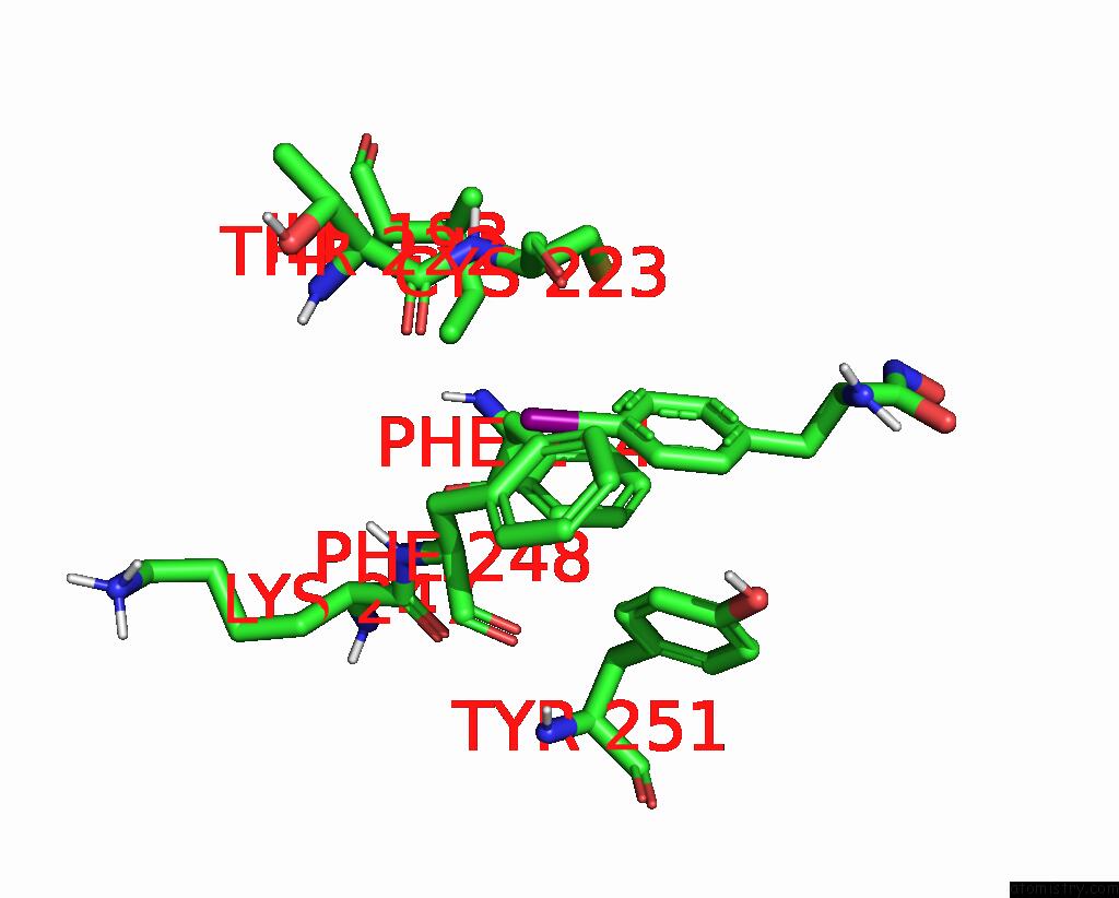
Mono view
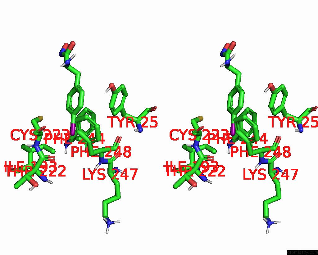
Stereo pair view
Mono view
Stereo pair view
A full contact list of Iodine with other atoms in the I binding
site number 1 of Aeromonas Proteolytica Aminopeptidase Complexed with the Inhibitor Para-Iodo-D-Phenylalanine Hydroxamate within 5.0Å range:
|
Reference:
B.Chevrier,
H.D'orchymont,
C.Schalk,
C.Tarnus,
D.Moras.
The Structure of the Aeromonas Proteolytica Aminopeptidase Complexed with A Hydroxamate Inhibitor. Involvement in Catalysis of GLU151 and Two Zinc Ions of the Co-Catalytic Unit. Eur.J.Biochem. V. 237 393 1996.
ISSN: ISSN 0014-2956
PubMed: 8647077
DOI: 10.1111/J.1432-1033.1996.0393K.X
Page generated: Fri Aug 8 11:57:24 2025
ISSN: ISSN 0014-2956
PubMed: 8647077
DOI: 10.1111/J.1432-1033.1996.0393K.X
Last articles
K in 5G0IK in 5FW3
K in 5G0J
K in 5G0R
K in 5FW2
K in 5G0H
K in 5FW1
K in 5FW5
K in 5FQ5
K in 5FUE