Iodine »
PDB 1gjd-1lij »
1ki6 »
Iodine in PDB 1ki6: Crystal Structure of Thymidine Kinase From Herpes Simplex Virus Type I Complexed with A 5-Iodouracil Anhydrohexitol Nucleoside
Enzymatic activity of Crystal Structure of Thymidine Kinase From Herpes Simplex Virus Type I Complexed with A 5-Iodouracil Anhydrohexitol Nucleoside
All present enzymatic activity of Crystal Structure of Thymidine Kinase From Herpes Simplex Virus Type I Complexed with A 5-Iodouracil Anhydrohexitol Nucleoside:
2.7.1.21;
2.7.1.21;
Protein crystallography data
The structure of Crystal Structure of Thymidine Kinase From Herpes Simplex Virus Type I Complexed with A 5-Iodouracil Anhydrohexitol Nucleoside, PDB code: 1ki6
was solved by
J.N.Champness,
M.S.Bennett,
F.Wien,
P.Herdewijn,
T.Ostrowski,
W.C.Summers,
M.R.Sanderson,
with X-Ray Crystallography technique. A brief refinement statistics is given in the table below:
| Resolution Low / High (Å) | 12.00 / 2.37 |
| Space group | C 2 2 21 |
| Cell size a, b, c (Å), α, β, γ (°) | 113.600, 116.400, 108.400, 90.00, 90.00, 90.00 |
| R / Rfree (%) | 22.3 / 31.7 |
Iodine Binding Sites:
The binding sites of Iodine atom in the Crystal Structure of Thymidine Kinase From Herpes Simplex Virus Type I Complexed with A 5-Iodouracil Anhydrohexitol Nucleoside
(pdb code 1ki6). This binding sites where shown within
5.0 Angstroms radius around Iodine atom.
In total 2 binding sites of Iodine where determined in the Crystal Structure of Thymidine Kinase From Herpes Simplex Virus Type I Complexed with A 5-Iodouracil Anhydrohexitol Nucleoside, PDB code: 1ki6:
Jump to Iodine binding site number: 1; 2;
In total 2 binding sites of Iodine where determined in the Crystal Structure of Thymidine Kinase From Herpes Simplex Virus Type I Complexed with A 5-Iodouracil Anhydrohexitol Nucleoside, PDB code: 1ki6:
Jump to Iodine binding site number: 1; 2;
Iodine binding site 1 out of 2 in 1ki6
Go back to
Iodine binding site 1 out
of 2 in the Crystal Structure of Thymidine Kinase From Herpes Simplex Virus Type I Complexed with A 5-Iodouracil Anhydrohexitol Nucleoside
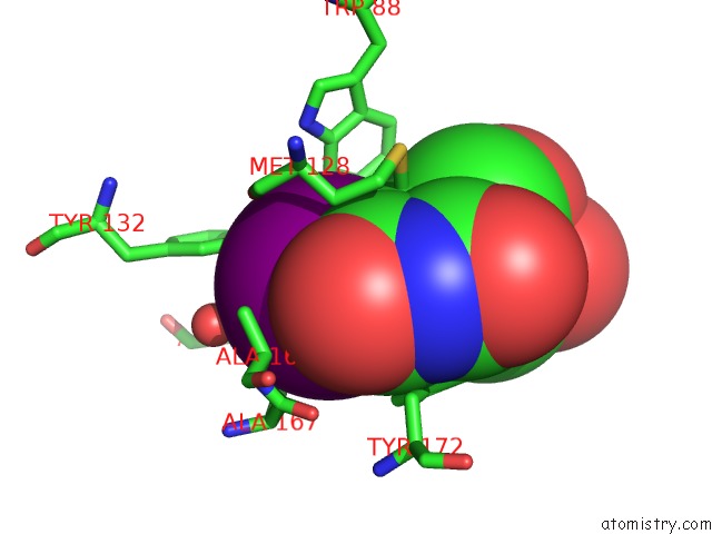
Mono view
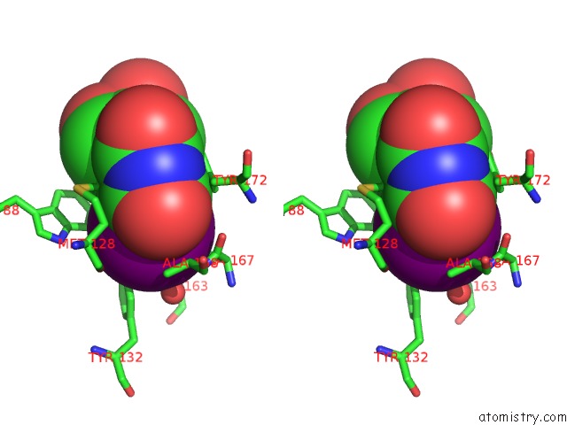
Stereo pair view
Mono view
Stereo pair view
A full contact list of Iodine with other atoms in the I binding
site number 1 of Crystal Structure of Thymidine Kinase From Herpes Simplex Virus Type I Complexed with A 5-Iodouracil Anhydrohexitol Nucleoside within 5.0Å range:
|
Iodine binding site 2 out of 2 in 1ki6
Go back to
Iodine binding site 2 out
of 2 in the Crystal Structure of Thymidine Kinase From Herpes Simplex Virus Type I Complexed with A 5-Iodouracil Anhydrohexitol Nucleoside
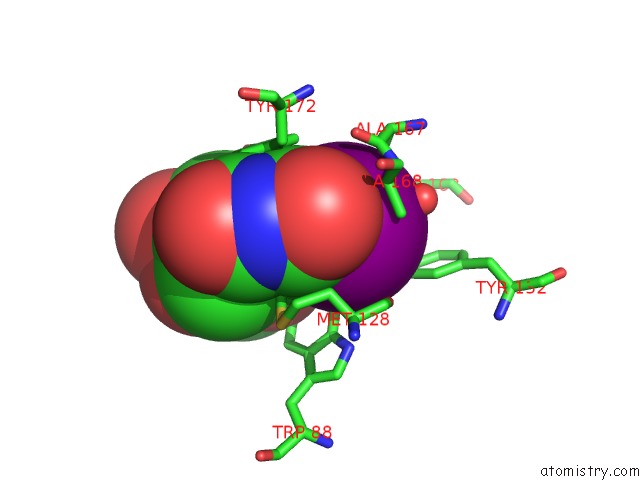
Mono view
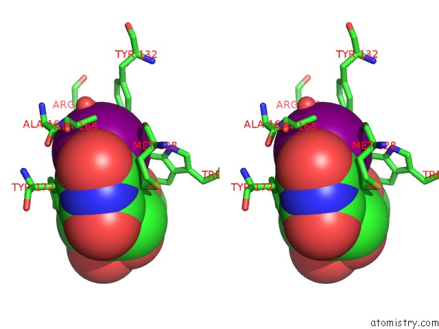
Stereo pair view
Mono view
Stereo pair view
A full contact list of Iodine with other atoms in the I binding
site number 2 of Crystal Structure of Thymidine Kinase From Herpes Simplex Virus Type I Complexed with A 5-Iodouracil Anhydrohexitol Nucleoside within 5.0Å range:
|
Reference:
J.N.Champness,
M.S.Bennett,
F.Wien,
R.Visse,
W.C.Summers,
P.Herdewijn,
E.De Clerq,
T.Ostrowski,
R.L.Jarvest,
M.R.Sanderson.
Exploring the Active Site of Herpes Simplex Virus Type-1 Thymidine Kinase By X-Ray Crystallography of Complexes with Aciclovir and Other Ligands. Proteins V. 32 350 1998.
ISSN: ISSN 0887-3585
PubMed: 9715911
DOI: 10.1002/(SICI)1097-0134(19980815)32:3<350::AID-PROT10>3.0.CO;2-8
Page generated: Fri Aug 8 11:59:29 2025
ISSN: ISSN 0887-3585
PubMed: 9715911
DOI: 10.1002/(SICI)1097-0134(19980815)32:3<350::AID-PROT10>3.0.CO;2-8
Last articles
Mg in 4DPGMg in 4DQP
Mg in 4DQQ
Mg in 4DPM
Mg in 4DPV
Mg in 4DQI
Mg in 4DOB
Mg in 4DOC
Mg in 4DMZ
Mg in 4DOA