Iodine »
PDB 1lkr-1pvh »
1mxi »
Iodine in PDB 1mxi: Structure of Yibk From Haemophilus Influenzae (HI0766): A Methyltransferase with A Cofactor Bound at A Site Formed By A Knot
Protein crystallography data
The structure of Structure of Yibk From Haemophilus Influenzae (HI0766): A Methyltransferase with A Cofactor Bound at A Site Formed By A Knot, PDB code: 1mxi
was solved by
K.Lim,
H.Zhang,
A.Tempczyk,
N.Bonander,
J.Toedt,
A.Howard,
E.Eisenstein,
O.Herzberg,
Structure 2 Function Project (S2F),
with X-Ray Crystallography technique. A brief refinement statistics is given in the table below:
| Resolution Low / High (Å) | 20.00 / 1.70 |
| Space group | P 41 21 2 |
| Cell size a, b, c (Å), α, β, γ (°) | 40.800, 40.800, 165.800, 90.00, 90.00, 90.00 |
| R / Rfree (%) | 19.6 / 25.4 |
Iodine Binding Sites:
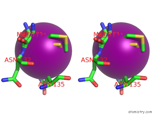
The binding sites of Iodine atom in the Structure of Yibk From Haemophilus Influenzae (HI0766): A Methyltransferase with A Cofactor Bound at A Site Formed By A Knot
(pdb code 1mxi). This binding sites where shown within
5.0 Angstroms radius around Iodine atom.
In total only one binding site of Iodine was determined in the Structure of Yibk From Haemophilus Influenzae (HI0766): A Methyltransferase with A Cofactor Bound at A Site Formed By A Knot, PDB code: 1mxi:
In total only one binding site of Iodine was determined in the Structure of Yibk From Haemophilus Influenzae (HI0766): A Methyltransferase with A Cofactor Bound at A Site Formed By A Knot, PDB code: 1mxi:
Iodine binding site 1 out of 1 in 1mxi
Go back to
Iodine binding site 1 out
of 1 in the Structure of Yibk From Haemophilus Influenzae (HI0766): A Methyltransferase with A Cofactor Bound at A Site Formed By A Knot

Mono view
Stereo pair view
Mono view
Stereo pair view
A full contact list of Iodine with other atoms in the I binding
site number 1 of Structure of Yibk From Haemophilus Influenzae (HI0766): A Methyltransferase with A Cofactor Bound at A Site Formed By A Knot within 5.0Å range:
|
Reference:
K.Lim,
H.Zhang,
A.Tempczyk,
W.Krajewski,
N.Bonander,
J.Toedt,
A.Howard,
E.Eisenstein,
O.Herzberg.
Structure of the Yibk Methyltransferase From Haemophilus Influenzae (HI0766): A Cofactor Bound at A Site Formed By A Knot Proteins V. 51 56 2003.
ISSN: ISSN 0887-3585
PubMed: 12596263
DOI: 10.1002/PROT.10323
Page generated: Sun Aug 11 12:28:50 2024
ISSN: ISSN 0887-3585
PubMed: 12596263
DOI: 10.1002/PROT.10323
Last articles
Zn in 9J0NZn in 9J0O
Zn in 9J0P
Zn in 9FJX
Zn in 9EKB
Zn in 9C0F
Zn in 9CAH
Zn in 9CH0
Zn in 9CH3
Zn in 9CH1