Iodine »
PDB 1lkr-1pvh »
1n4g »
Iodine in PDB 1n4g: Structure of CYP121, A Mycobacterial P450, in Complex with Iodopyrazole
Protein crystallography data
The structure of Structure of CYP121, A Mycobacterial P450, in Complex with Iodopyrazole, PDB code: 1n4g
was solved by
D.Leys,
C.G.Mowat,
K.J.Mclean,
A.Richmond,
S.K.Chapman,
M.D.Walkinshaw,
A.W.Munro,
Tb Structural Genomics Consortium(Tbsgc),
with X-Ray Crystallography technique. A brief refinement statistics is given in the table below:
| Resolution Low / High (Å) | 9.98 / 1.80 |
| Space group | P 65 2 2 |
| Cell size a, b, c (Å), α, β, γ (°) | 78.717, 78.717, 268.240, 90.00, 90.00, 120.00 |
| R / Rfree (%) | 20.5 / 21.7 |
Other elements in 1n4g:
The structure of Structure of CYP121, A Mycobacterial P450, in Complex with Iodopyrazole also contains other interesting chemical elements:
| Iron | (Fe) | 1 atom |
Iodine Binding Sites:
The binding sites of Iodine atom in the Structure of CYP121, A Mycobacterial P450, in Complex with Iodopyrazole
(pdb code 1n4g). This binding sites where shown within
5.0 Angstroms radius around Iodine atom.
In total only one binding site of Iodine was determined in the Structure of CYP121, A Mycobacterial P450, in Complex with Iodopyrazole, PDB code: 1n4g:
In total only one binding site of Iodine was determined in the Structure of CYP121, A Mycobacterial P450, in Complex with Iodopyrazole, PDB code: 1n4g:
Iodine binding site 1 out of 1 in 1n4g
Go back to
Iodine binding site 1 out
of 1 in the Structure of CYP121, A Mycobacterial P450, in Complex with Iodopyrazole
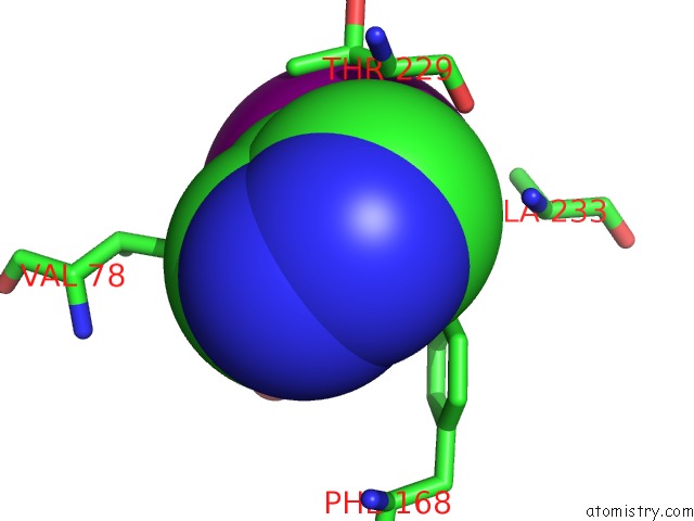
Mono view
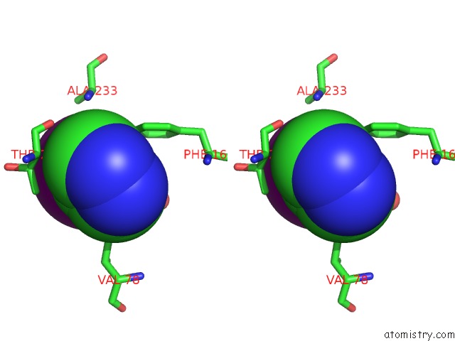
Stereo pair view
Mono view
Stereo pair view
A full contact list of Iodine with other atoms in the I binding
site number 1 of Structure of CYP121, A Mycobacterial P450, in Complex with Iodopyrazole within 5.0Å range:
|
Reference:
D.Leys,
C.G.Mowat,
K.J.Mclean,
A.Richmond,
S.K.Chapman,
M.D.Walkinshaw,
A.W.Munro.
Atomic Structure of Mycobacterium Tuberculosis CYP121 to 1.06 A Reveals Novel Features of Cytochrome P450. J.Biol.Chem. V. 278 5141 2003.
ISSN: ISSN 0021-9258
PubMed: 12435731
DOI: 10.1074/JBC.M209928200
Page generated: Sun Aug 11 12:29:20 2024
ISSN: ISSN 0021-9258
PubMed: 12435731
DOI: 10.1074/JBC.M209928200
Last articles
Zn in 9J0NZn in 9J0O
Zn in 9J0P
Zn in 9FJX
Zn in 9EKB
Zn in 9C0F
Zn in 9CAH
Zn in 9CH0
Zn in 9CH3
Zn in 9CH1