Iodine »
PDB 1lkr-1pvh »
1orw »
Iodine in PDB 1orw: Crystal Structure of Porcine Dipeptidyl Peptidase IV (CD26) in Complex with A Peptidomimetic Inhibitor
Enzymatic activity of Crystal Structure of Porcine Dipeptidyl Peptidase IV (CD26) in Complex with A Peptidomimetic Inhibitor
All present enzymatic activity of Crystal Structure of Porcine Dipeptidyl Peptidase IV (CD26) in Complex with A Peptidomimetic Inhibitor:
3.4.14.5;
3.4.14.5;
Protein crystallography data
The structure of Crystal Structure of Porcine Dipeptidyl Peptidase IV (CD26) in Complex with A Peptidomimetic Inhibitor, PDB code: 1orw
was solved by
M.Engel,
T.Hoffmann,
L.Wagner,
M.Wermann,
U.Heiser,
R.Kiefersauer,
R.Huber,
W.Bode,
H.U.Demuth,
H.Brandstetter,
with X-Ray Crystallography technique. A brief refinement statistics is given in the table below:
| Resolution Low / High (Å) | 29.79 / 2.84 |
| Space group | P 1 |
| Cell size a, b, c (Å), α, β, γ (°) | 61.970, 117.710, 133.560, 112.65, 94.81, 91.17 |
| R / Rfree (%) | 18.7 / 24.6 |
Iodine Binding Sites:
The binding sites of Iodine atom in the Crystal Structure of Porcine Dipeptidyl Peptidase IV (CD26) in Complex with A Peptidomimetic Inhibitor
(pdb code 1orw). This binding sites where shown within
5.0 Angstroms radius around Iodine atom.
In total 4 binding sites of Iodine where determined in the Crystal Structure of Porcine Dipeptidyl Peptidase IV (CD26) in Complex with A Peptidomimetic Inhibitor, PDB code: 1orw:
Jump to Iodine binding site number: 1; 2; 3; 4;
In total 4 binding sites of Iodine where determined in the Crystal Structure of Porcine Dipeptidyl Peptidase IV (CD26) in Complex with A Peptidomimetic Inhibitor, PDB code: 1orw:
Jump to Iodine binding site number: 1; 2; 3; 4;
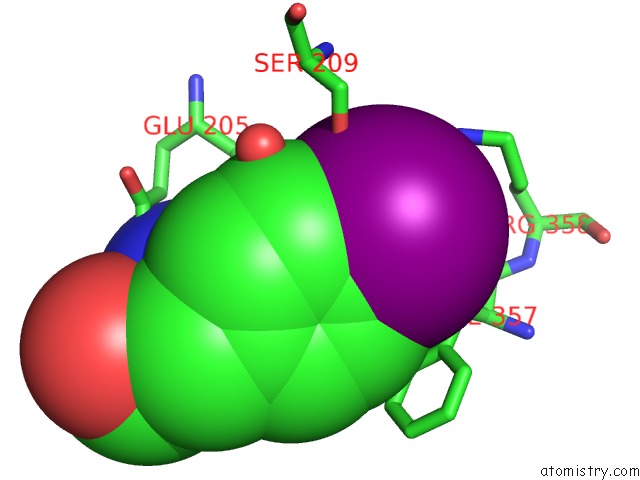
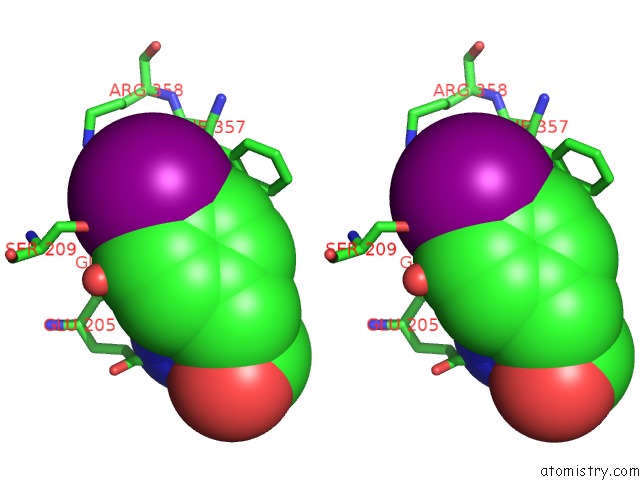
Iodine binding site 1 out of 4 in 1orw
Go back to
Iodine binding site 1 out
of 4 in the Crystal Structure of Porcine Dipeptidyl Peptidase IV (CD26) in Complex with A Peptidomimetic Inhibitor
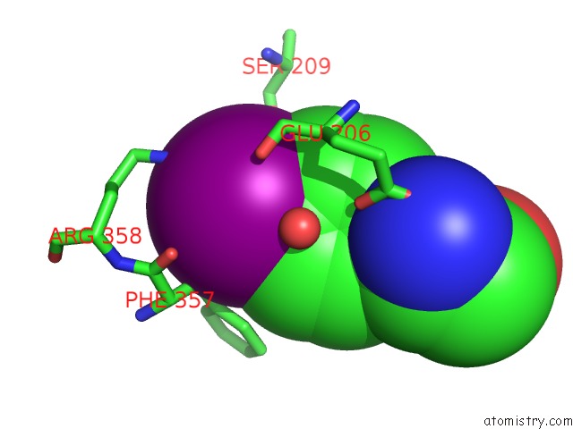
Mono view
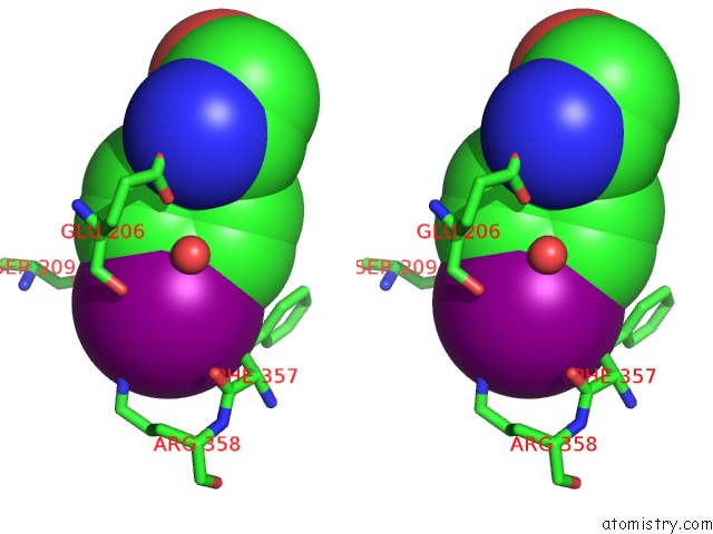
Stereo pair view
Mono view
Stereo pair view
A full contact list of Iodine with other atoms in the I binding
site number 1 of Crystal Structure of Porcine Dipeptidyl Peptidase IV (CD26) in Complex with A Peptidomimetic Inhibitor within 5.0Å range:
|
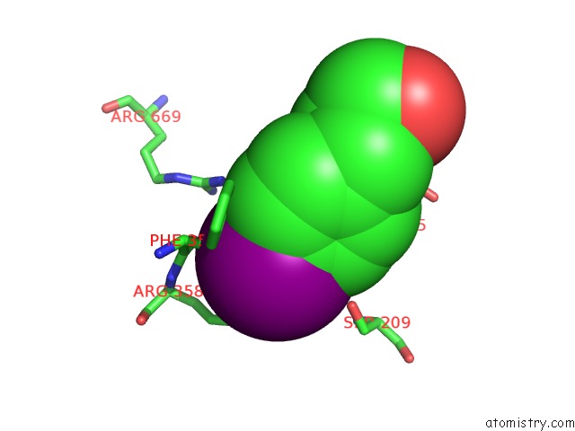
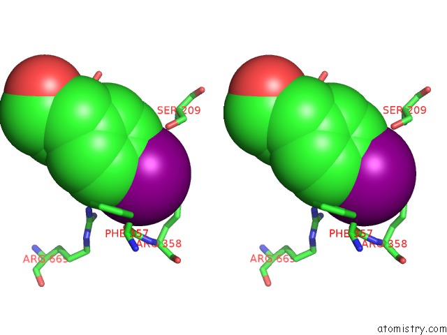
Iodine binding site 2 out of 4 in 1orw
Go back to
Iodine binding site 2 out
of 4 in the Crystal Structure of Porcine Dipeptidyl Peptidase IV (CD26) in Complex with A Peptidomimetic Inhibitor
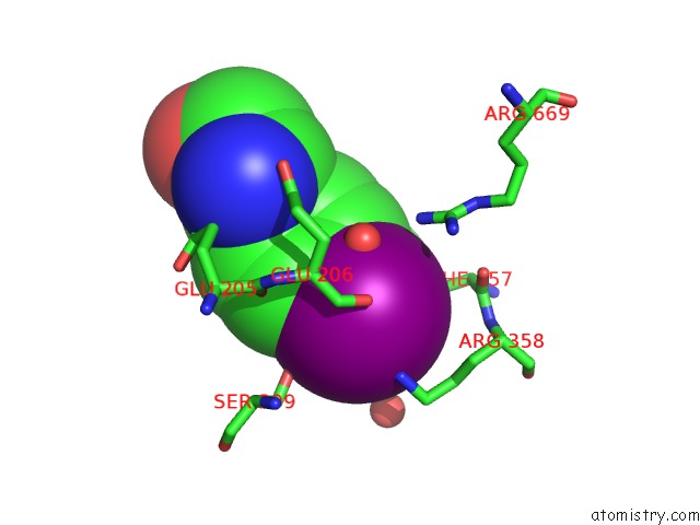
Mono view
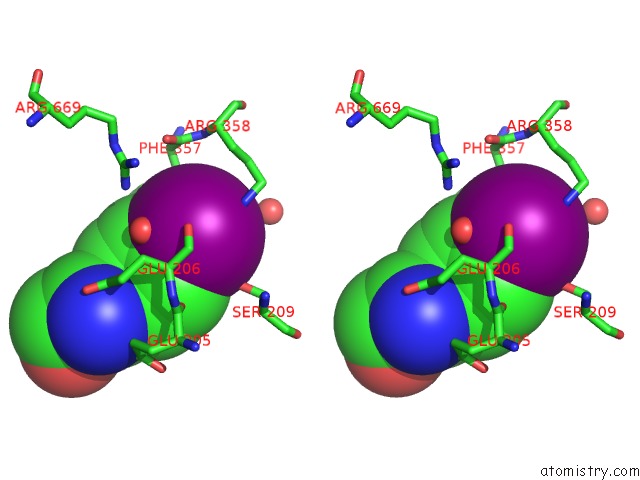
Stereo pair view
Mono view
Stereo pair view
A full contact list of Iodine with other atoms in the I binding
site number 2 of Crystal Structure of Porcine Dipeptidyl Peptidase IV (CD26) in Complex with A Peptidomimetic Inhibitor within 5.0Å range:
|
Iodine binding site 3 out of 4 in 1orw
Go back to
Iodine binding site 3 out
of 4 in the Crystal Structure of Porcine Dipeptidyl Peptidase IV (CD26) in Complex with A Peptidomimetic Inhibitor
Mono view
Stereo pair view
Mono view
Stereo pair view
A full contact list of Iodine with other atoms in the I binding
site number 3 of Crystal Structure of Porcine Dipeptidyl Peptidase IV (CD26) in Complex with A Peptidomimetic Inhibitor within 5.0Å range:
|
Iodine binding site 4 out of 4 in 1orw
Go back to
Iodine binding site 4 out
of 4 in the Crystal Structure of Porcine Dipeptidyl Peptidase IV (CD26) in Complex with A Peptidomimetic Inhibitor
Mono view
Stereo pair view
Mono view
Stereo pair view
A full contact list of Iodine with other atoms in the I binding
site number 4 of Crystal Structure of Porcine Dipeptidyl Peptidase IV (CD26) in Complex with A Peptidomimetic Inhibitor within 5.0Å range:
|
Reference:
M.Engel,
T.Hoffmann,
L.Wagner,
M.Wermann,
U.Heiser,
R.Kiefersauer,
R.Huber,
W.Bode,
H.U.Demuth,
H.Brandstetter.
The Crystal Structure of Dipeptidyl Peptidase IV (CD26) Reveals Its Functional Regulation and Enzymatic Mechanism Proc.Natl.Acad.Sci.Usa V. 100 5063 2003.
ISSN: ISSN 0027-8424
PubMed: 12690074
DOI: 10.1073/PNAS.0230620100
Page generated: Sun Aug 11 12:33:34 2024
ISSN: ISSN 0027-8424
PubMed: 12690074
DOI: 10.1073/PNAS.0230620100
Last articles
Zn in 9J0NZn in 9J0O
Zn in 9J0P
Zn in 9FJX
Zn in 9EKB
Zn in 9C0F
Zn in 9CAH
Zn in 9CH0
Zn in 9CH3
Zn in 9CH1