Iodine »
PDB 1lkr-1pvh »
1pvh »
Iodine in PDB 1pvh: Crystal Structure of Leukemia Inhibitory Factor in Complex with GP130
Protein crystallography data
The structure of Crystal Structure of Leukemia Inhibitory Factor in Complex with GP130, PDB code: 1pvh
was solved by
M.J.Boulanger,
A.J.Bankovich,
T.Kortemme,
D.Baker,
K.C.Garcia,
with X-Ray Crystallography technique. A brief refinement statistics is given in the table below:
| Resolution Low / High (Å) | 39.86 / 2.50 |
| Space group | P 21 21 21 |
| Cell size a, b, c (Å), α, β, γ (°) | 79.710, 86.700, 146.430, 90.00, 90.00, 90.00 |
| R / Rfree (%) | 24.8 / 28.9 |
Iodine Binding Sites:
The binding sites of Iodine atom in the Crystal Structure of Leukemia Inhibitory Factor in Complex with GP130
(pdb code 1pvh). This binding sites where shown within
5.0 Angstroms radius around Iodine atom.
In total 2 binding sites of Iodine where determined in the Crystal Structure of Leukemia Inhibitory Factor in Complex with GP130, PDB code: 1pvh:
Jump to Iodine binding site number: 1; 2;
In total 2 binding sites of Iodine where determined in the Crystal Structure of Leukemia Inhibitory Factor in Complex with GP130, PDB code: 1pvh:
Jump to Iodine binding site number: 1; 2;
Iodine binding site 1 out of 2 in 1pvh
Go back to
Iodine binding site 1 out
of 2 in the Crystal Structure of Leukemia Inhibitory Factor in Complex with GP130
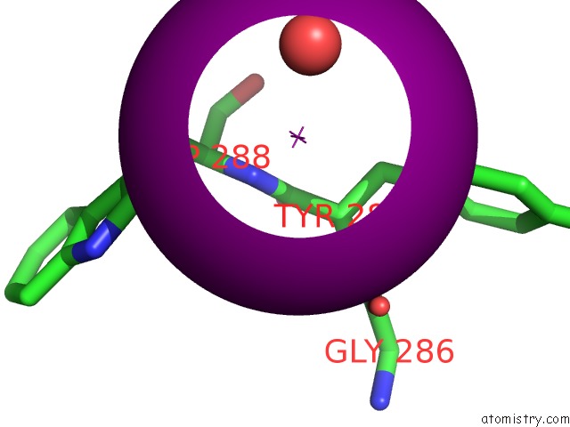
Mono view
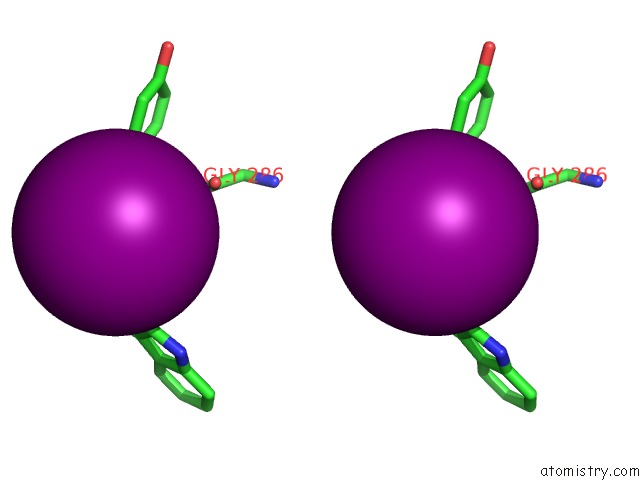
Stereo pair view
Mono view
Stereo pair view
A full contact list of Iodine with other atoms in the I binding
site number 1 of Crystal Structure of Leukemia Inhibitory Factor in Complex with GP130 within 5.0Å range:
|
Iodine binding site 2 out of 2 in 1pvh
Go back to
Iodine binding site 2 out
of 2 in the Crystal Structure of Leukemia Inhibitory Factor in Complex with GP130
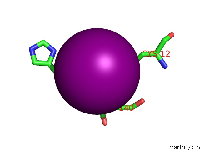
Mono view
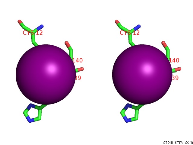
Stereo pair view
Mono view
Stereo pair view
A full contact list of Iodine with other atoms in the I binding
site number 2 of Crystal Structure of Leukemia Inhibitory Factor in Complex with GP130 within 5.0Å range:
|
Reference:
M.J.Boulanger,
A.J.Bankovich,
T.Kortemme,
D.Baker,
K.C.Garcia.
Convergent Mechanisms For Recognition of Divergent Cytokines By the Shared Signaling Receptor GP130. Mol.Cell V. 12 577 2003.
ISSN: ISSN 1097-2765
PubMed: 14527405
DOI: 10.1016/S1097-2765(03)00365-4
Page generated: Sun Aug 11 12:37:20 2024
ISSN: ISSN 1097-2765
PubMed: 14527405
DOI: 10.1016/S1097-2765(03)00365-4
Last articles
Zn in 9J0NZn in 9J0O
Zn in 9J0P
Zn in 9FJX
Zn in 9EKB
Zn in 9C0F
Zn in 9CAH
Zn in 9CH0
Zn in 9CH3
Zn in 9CH1