Iodine »
PDB 1q0t-1v1f »
1tb4 »
Iodine in PDB 1tb4: Crystal Structure of Aspartate-Semialdehyde Dehydrogenase From Haemophilus Influenzae with A Bound Periodate
Enzymatic activity of Crystal Structure of Aspartate-Semialdehyde Dehydrogenase From Haemophilus Influenzae with A Bound Periodate
All present enzymatic activity of Crystal Structure of Aspartate-Semialdehyde Dehydrogenase From Haemophilus Influenzae with A Bound Periodate:
1.2.1.11;
1.2.1.11;
Protein crystallography data
The structure of Crystal Structure of Aspartate-Semialdehyde Dehydrogenase From Haemophilus Influenzae with A Bound Periodate, PDB code: 1tb4
was solved by
R.E.Viola,
with X-Ray Crystallography technique. A brief refinement statistics is given in the table below:
| Resolution Low / High (Å) | 50.00 / 2.15 |
| Space group | P 21 21 2 |
| Cell size a, b, c (Å), α, β, γ (°) | 113.999, 55.146, 57.646, 90.00, 90.00, 90.00 |
| R / Rfree (%) | 23 / 28.8 |
Iodine Binding Sites:
The binding sites of Iodine atom in the Crystal Structure of Aspartate-Semialdehyde Dehydrogenase From Haemophilus Influenzae with A Bound Periodate
(pdb code 1tb4). This binding sites where shown within
5.0 Angstroms radius around Iodine atom.
In total only one binding site of Iodine was determined in the Crystal Structure of Aspartate-Semialdehyde Dehydrogenase From Haemophilus Influenzae with A Bound Periodate, PDB code: 1tb4:
In total only one binding site of Iodine was determined in the Crystal Structure of Aspartate-Semialdehyde Dehydrogenase From Haemophilus Influenzae with A Bound Periodate, PDB code: 1tb4:
Iodine binding site 1 out of 1 in 1tb4
Go back to
Iodine binding site 1 out
of 1 in the Crystal Structure of Aspartate-Semialdehyde Dehydrogenase From Haemophilus Influenzae with A Bound Periodate
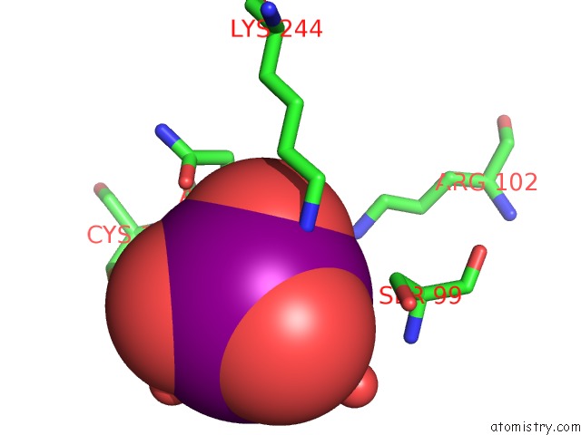
Mono view
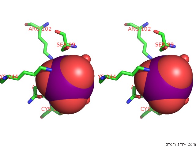
Stereo pair view
Mono view
Stereo pair view
A full contact list of Iodine with other atoms in the I binding
site number 1 of Crystal Structure of Aspartate-Semialdehyde Dehydrogenase From Haemophilus Influenzae with A Bound Periodate within 5.0Å range:
|
Reference:
C.R.Faehnle,
J.Blanco,
R.E.Viola.
Structural Basis For Discrimination Between Oxyanion Substrates or Inhibitors in Aspartate-Beta-Semialdehyde Dehydrogenase. Acta Crystallogr.,Sect.D V. 60 2320 2004.
ISSN: ISSN 0907-4449
PubMed: 15583380
DOI: 10.1107/S0907444904026411
Page generated: Sun Aug 11 12:52:00 2024
ISSN: ISSN 0907-4449
PubMed: 15583380
DOI: 10.1107/S0907444904026411
Last articles
Zn in 9J0NZn in 9J0O
Zn in 9J0P
Zn in 9FJX
Zn in 9EKB
Zn in 9C0F
Zn in 9CAH
Zn in 9CH0
Zn in 9CH3
Zn in 9CH1