Iodine »
PDB 1v1g-2arl »
1v3o »
Iodine in PDB 1v3o: Crystal Structure of D(Gcgagagc): the Dna Quadruplex Structure Split From the Octaplex
Protein crystallography data
The structure of Crystal Structure of D(Gcgagagc): the Dna Quadruplex Structure Split From the Octaplex, PDB code: 1v3o
was solved by
J.Kondo,
S.Umeda,
T.Sunami,
A.Takenaka,
with X-Ray Crystallography technique. A brief refinement statistics is given in the table below:
| Resolution Low / High (Å) | 9.00 / 1.70 |
| Space group | I 2 2 2 |
| Cell size a, b, c (Å), α, β, γ (°) | 34.650, 42.490, 64.080, 90.00, 90.00, 90.00 |
| R / Rfree (%) | 26.2 / 29.6 |
Other elements in 1v3o:
The structure of Crystal Structure of D(Gcgagagc): the Dna Quadruplex Structure Split From the Octaplex also contains other interesting chemical elements:
| Potassium | (K) | 1 atom |
Iodine Binding Sites:
The binding sites of Iodine atom in the Crystal Structure of D(Gcgagagc): the Dna Quadruplex Structure Split From the Octaplex
(pdb code 1v3o). This binding sites where shown within
5.0 Angstroms radius around Iodine atom.
In total 2 binding sites of Iodine where determined in the Crystal Structure of D(Gcgagagc): the Dna Quadruplex Structure Split From the Octaplex, PDB code: 1v3o:
Jump to Iodine binding site number: 1; 2;
In total 2 binding sites of Iodine where determined in the Crystal Structure of D(Gcgagagc): the Dna Quadruplex Structure Split From the Octaplex, PDB code: 1v3o:
Jump to Iodine binding site number: 1; 2;
Iodine binding site 1 out of 2 in 1v3o
Go back to
Iodine binding site 1 out
of 2 in the Crystal Structure of D(Gcgagagc): the Dna Quadruplex Structure Split From the Octaplex
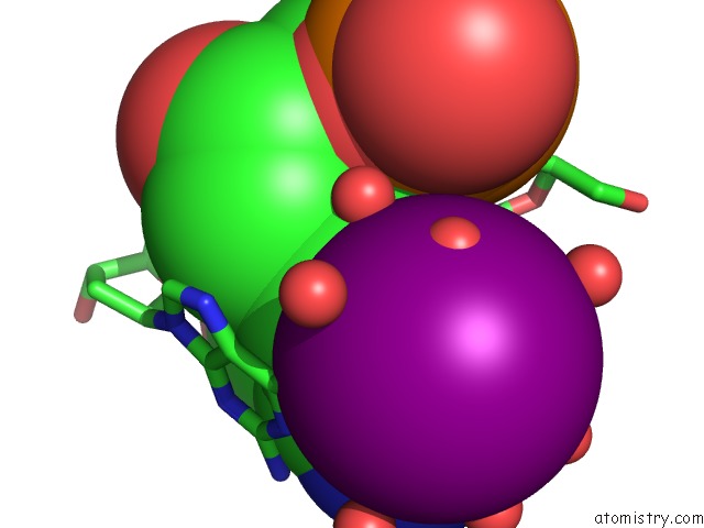
Mono view
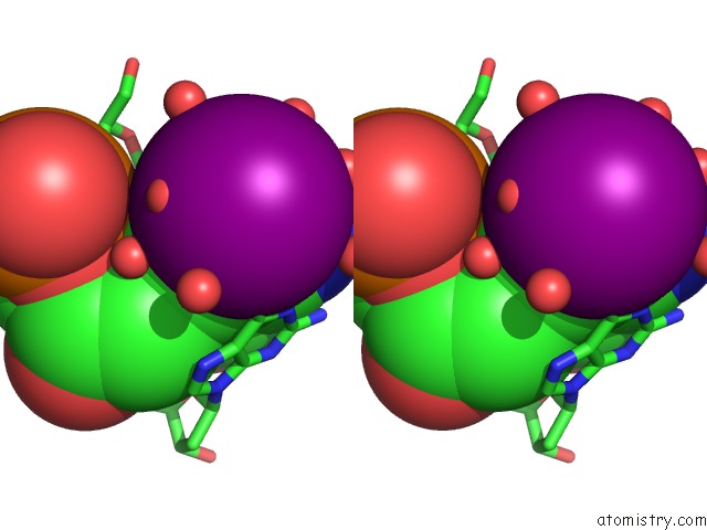
Stereo pair view
Mono view
Stereo pair view
A full contact list of Iodine with other atoms in the I binding
site number 1 of Crystal Structure of D(Gcgagagc): the Dna Quadruplex Structure Split From the Octaplex within 5.0Å range:
|
Iodine binding site 2 out of 2 in 1v3o
Go back to
Iodine binding site 2 out
of 2 in the Crystal Structure of D(Gcgagagc): the Dna Quadruplex Structure Split From the Octaplex
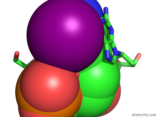
Mono view
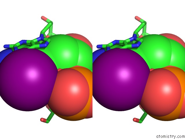
Stereo pair view
Mono view
Stereo pair view
A full contact list of Iodine with other atoms in the I binding
site number 2 of Crystal Structure of D(Gcgagagc): the Dna Quadruplex Structure Split From the Octaplex within 5.0Å range:
|
Reference:
J.Kondo,
W.Adachi,
S.Umeda,
T.Sunami,
A.Takenaka.
Crystal Structures of A Dna Octaplex with I-Motif of G-Quartets and Its Splitting Into Two Quadruplexes Suggest A Folding Mechanism of Eight Tandem Repeats Nucleic Acids Res. V. 32 2541 2004.
ISSN: ISSN 0305-1048
PubMed: 15133122
DOI: 10.1093/NAR/GKH575
Page generated: Sun Aug 11 12:58:43 2024
ISSN: ISSN 0305-1048
PubMed: 15133122
DOI: 10.1093/NAR/GKH575
Last articles
Zn in 9J0NZn in 9J0O
Zn in 9J0P
Zn in 9FJX
Zn in 9EKB
Zn in 9C0F
Zn in 9CAH
Zn in 9CH0
Zn in 9CH3
Zn in 9CH1