Iodine »
PDB 1v1g-2arl »
1vj5 »
Iodine in PDB 1vj5: Human Soluble Epoxide Hydrolase- N-Cyclohexyl-N'-(4-Iodophenyl)Urea Complex
Enzymatic activity of Human Soluble Epoxide Hydrolase- N-Cyclohexyl-N'-(4-Iodophenyl)Urea Complex
All present enzymatic activity of Human Soluble Epoxide Hydrolase- N-Cyclohexyl-N'-(4-Iodophenyl)Urea Complex:
3.3.2.3;
3.3.2.3;
Protein crystallography data
The structure of Human Soluble Epoxide Hydrolase- N-Cyclohexyl-N'-(4-Iodophenyl)Urea Complex, PDB code: 1vj5
was solved by
G.A.Gomez,
C.Morisseau,
B.D.Hammock,
D.W.Christianson,
with X-Ray Crystallography technique. A brief refinement statistics is given in the table below:
| Resolution Low / High (Å) | 48.94 / 2.35 |
| Space group | P 65 2 2 |
| Cell size a, b, c (Å), α, β, γ (°) | 93.510, 93.510, 245.700, 90.00, 90.00, 120.00 |
| R / Rfree (%) | 21.7 / 26.6 |
Other elements in 1vj5:
The structure of Human Soluble Epoxide Hydrolase- N-Cyclohexyl-N'-(4-Iodophenyl)Urea Complex also contains other interesting chemical elements:
| Magnesium | (Mg) | 1 atom |
Iodine Binding Sites:
The binding sites of Iodine atom in the Human Soluble Epoxide Hydrolase- N-Cyclohexyl-N'-(4-Iodophenyl)Urea Complex
(pdb code 1vj5). This binding sites where shown within
5.0 Angstroms radius around Iodine atom.
In total only one binding site of Iodine was determined in the Human Soluble Epoxide Hydrolase- N-Cyclohexyl-N'-(4-Iodophenyl)Urea Complex, PDB code: 1vj5:
In total only one binding site of Iodine was determined in the Human Soluble Epoxide Hydrolase- N-Cyclohexyl-N'-(4-Iodophenyl)Urea Complex, PDB code: 1vj5:
Iodine binding site 1 out of 1 in 1vj5
Go back to
Iodine binding site 1 out
of 1 in the Human Soluble Epoxide Hydrolase- N-Cyclohexyl-N'-(4-Iodophenyl)Urea Complex
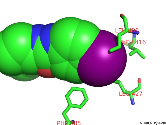
Mono view
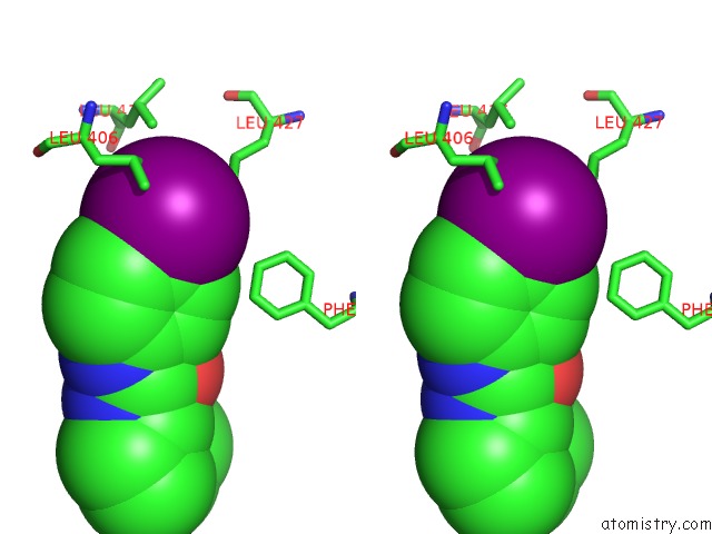
Stereo pair view
Mono view
Stereo pair view
A full contact list of Iodine with other atoms in the I binding
site number 1 of Human Soluble Epoxide Hydrolase- N-Cyclohexyl-N'-(4-Iodophenyl)Urea Complex within 5.0Å range:
|
Reference:
G.A.Gomez,
C.Morisseau,
B.D.Hammock,
D.W.Christianson.
Structure of Human Epoxide Hydrolase Reveals Mechanistic Inferences on Bifunctional Catalysis in Epoxide and Phosphate Ester Hydrolysis Biochemistry V. 43 4716 2004.
ISSN: ISSN 0006-2960
PubMed: 15096040
DOI: 10.1021/BI036189J
Page generated: Sun Aug 11 12:58:43 2024
ISSN: ISSN 0006-2960
PubMed: 15096040
DOI: 10.1021/BI036189J
Last articles
Zn in 9MJ5Zn in 9HNW
Zn in 9G0L
Zn in 9FNE
Zn in 9DZN
Zn in 9E0I
Zn in 9D32
Zn in 9DAK
Zn in 8ZXC
Zn in 8ZUF