Iodine »
PDB 1v1g-2arl »
2anv »
Iodine in PDB 2anv: Crystal Structure of P22 Lysozyme Mutant L86M
Enzymatic activity of Crystal Structure of P22 Lysozyme Mutant L86M
All present enzymatic activity of Crystal Structure of P22 Lysozyme Mutant L86M:
3.2.1.17;
3.2.1.17;
Protein crystallography data
The structure of Crystal Structure of P22 Lysozyme Mutant L86M, PDB code: 2anv
was solved by
B.H.Mooers,
B.W.Matthews,
with X-Ray Crystallography technique. A brief refinement statistics is given in the table below:
| Resolution Low / High (Å) | 42.00 / 1.04 |
| Space group | C 1 2 1 |
| Cell size a, b, c (Å), α, β, γ (°) | 134.013, 50.415, 46.587, 90.00, 103.80, 90.00 |
| R / Rfree (%) | 12.2 / 15.1 |
Other elements in 2anv:
The structure of Crystal Structure of P22 Lysozyme Mutant L86M also contains other interesting chemical elements:
| Magnesium | (Mg) | 1 atom |
| Samarium | (Sm) | 3 atoms |
| Chlorine | (Cl) | 3 atoms |
Iodine Binding Sites:
The binding sites of Iodine atom in the Crystal Structure of P22 Lysozyme Mutant L86M
(pdb code 2anv). This binding sites where shown within
5.0 Angstroms radius around Iodine atom.
In total 6 binding sites of Iodine where determined in the Crystal Structure of P22 Lysozyme Mutant L86M, PDB code: 2anv:
Jump to Iodine binding site number: 1; 2; 3; 4; 5; 6;
In total 6 binding sites of Iodine where determined in the Crystal Structure of P22 Lysozyme Mutant L86M, PDB code: 2anv:
Jump to Iodine binding site number: 1; 2; 3; 4; 5; 6;
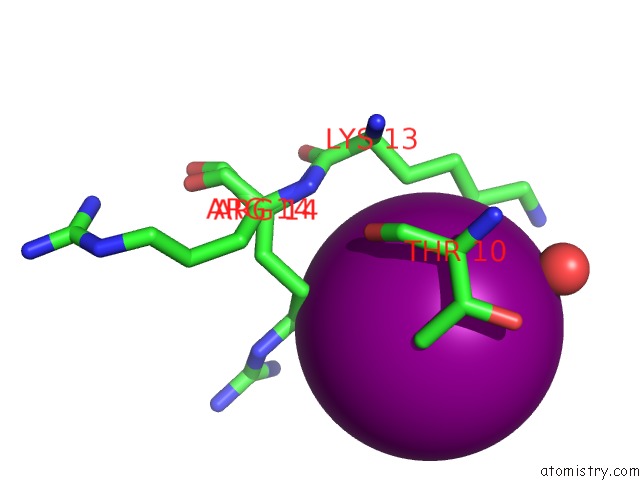
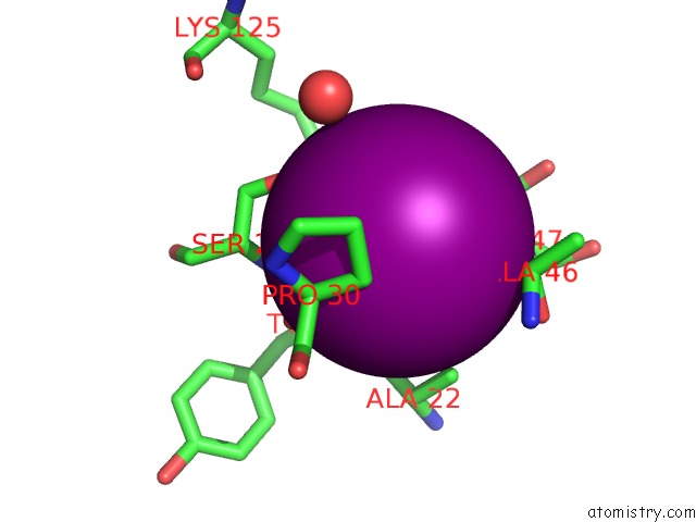
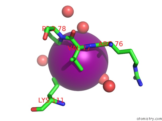
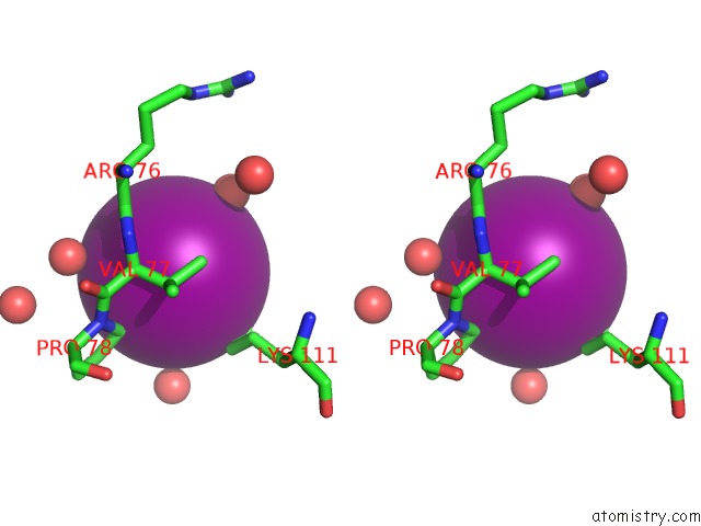
Iodine binding site 1 out of 6 in 2anv
Go back to
Iodine binding site 1 out
of 6 in the Crystal Structure of P22 Lysozyme Mutant L86M
Mono view
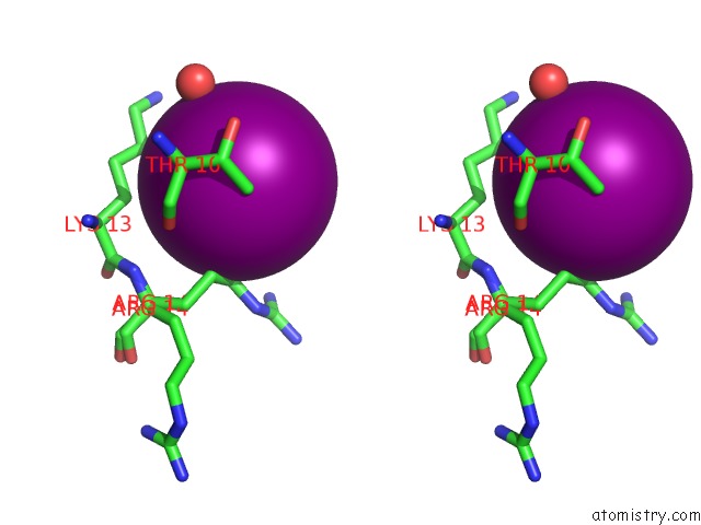
Stereo pair view
Mono view
Stereo pair view
A full contact list of Iodine with other atoms in the I binding
site number 1 of Crystal Structure of P22 Lysozyme Mutant L86M within 5.0Å range:
|
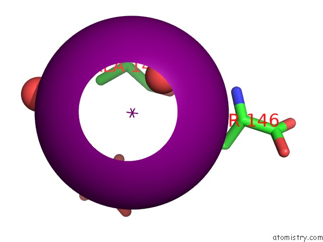
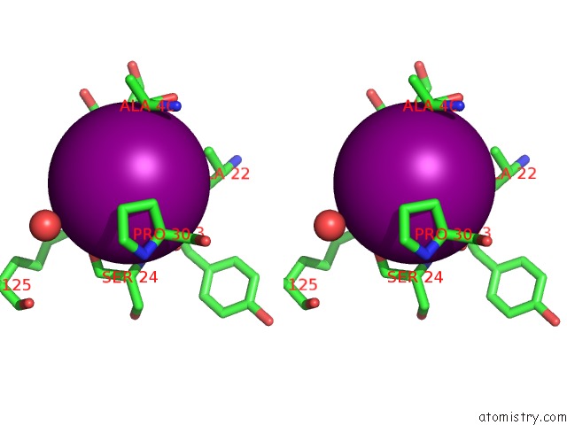
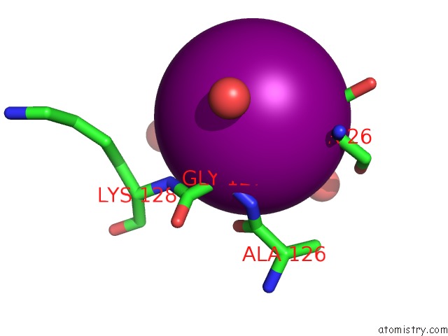
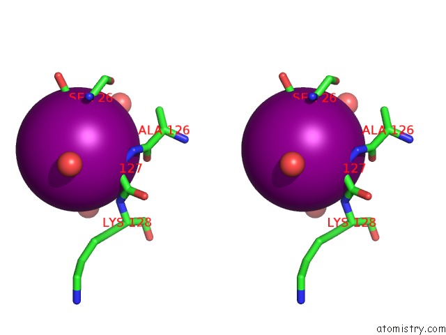
Iodine binding site 2 out of 6 in 2anv
Go back to
Iodine binding site 2 out
of 6 in the Crystal Structure of P22 Lysozyme Mutant L86M
Mono view
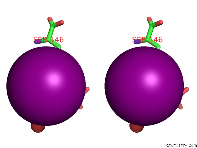
Stereo pair view
Mono view
Stereo pair view
A full contact list of Iodine with other atoms in the I binding
site number 2 of Crystal Structure of P22 Lysozyme Mutant L86M within 5.0Å range:
|
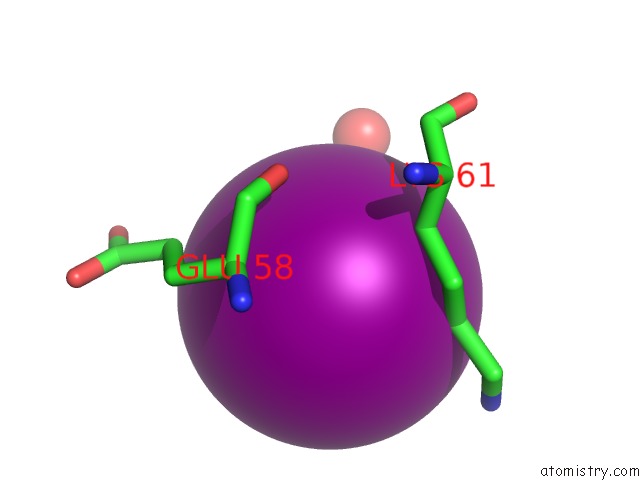
Iodine binding site 3 out of 6 in 2anv
Go back to
Iodine binding site 3 out
of 6 in the Crystal Structure of P22 Lysozyme Mutant L86M
Mono view
Stereo pair view
Mono view
Stereo pair view
A full contact list of Iodine with other atoms in the I binding
site number 3 of Crystal Structure of P22 Lysozyme Mutant L86M within 5.0Å range:
|
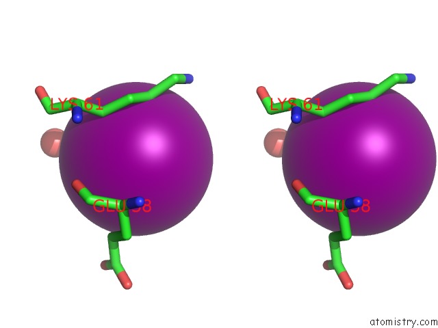
Iodine binding site 4 out of 6 in 2anv
Go back to
Iodine binding site 4 out
of 6 in the Crystal Structure of P22 Lysozyme Mutant L86M
Mono view
Stereo pair view
Mono view
Stereo pair view
A full contact list of Iodine with other atoms in the I binding
site number 4 of Crystal Structure of P22 Lysozyme Mutant L86M within 5.0Å range:
|
Iodine binding site 5 out of 6 in 2anv
Go back to
Iodine binding site 5 out
of 6 in the Crystal Structure of P22 Lysozyme Mutant L86M
Mono view
Stereo pair view
Mono view
Stereo pair view
A full contact list of Iodine with other atoms in the I binding
site number 5 of Crystal Structure of P22 Lysozyme Mutant L86M within 5.0Å range:
|
Iodine binding site 6 out of 6 in 2anv
Go back to
Iodine binding site 6 out
of 6 in the Crystal Structure of P22 Lysozyme Mutant L86M
Mono view
Stereo pair view
Mono view
Stereo pair view
A full contact list of Iodine with other atoms in the I binding
site number 6 of Crystal Structure of P22 Lysozyme Mutant L86M within 5.0Å range:
|
Reference:
B.H.Mooers,
B.W.Matthews.
Extension to 2268 Atoms of Direct Methods in the Ab Initio Determination of the Unknown Structure of Bacteriophage P22 Lysozyme. Acta Crystallogr.,Sect.D V. 62 165 2006.
ISSN: ISSN 0907-4449
PubMed: 16421448
DOI: 10.1107/S0907444905037212
Page generated: Sun Aug 11 13:14:07 2024
ISSN: ISSN 0907-4449
PubMed: 16421448
DOI: 10.1107/S0907444905037212
Last articles
Cl in 5UJBCl in 5UGS
Cl in 5UJS
Cl in 5UHI
Cl in 5UJK
Cl in 5UJ2
Cl in 5UIN
Cl in 5UI2
Cl in 5UGT
Cl in 5UHR