Iodine »
PDB 2axe-2fwh »
2ckl »
Iodine in PDB 2ckl: RING1B-BMI1 E3 Catalytic Domain Structure
Protein crystallography data
The structure of RING1B-BMI1 E3 Catalytic Domain Structure, PDB code: 2ckl
was solved by
G.Buchwald,
P.Van Der Stoop,
O.Weichenrieder,
A.Perrakis,
M.Van Lohuizen,
T.K.Sixma,
with X-Ray Crystallography technique. A brief refinement statistics is given in the table below:
| Resolution Low / High (Å) | 30.00 / 2.0 |
| Space group | P 63 |
| Cell size a, b, c (Å), α, β, γ (°) | 120.046, 120.046, 27.096, 90.00, 90.00, 120.00 |
| R / Rfree (%) | 17.9 / 21.3 |
Other elements in 2ckl:
The structure of RING1B-BMI1 E3 Catalytic Domain Structure also contains other interesting chemical elements:
| Zinc | (Zn) | 4 atoms |
Iodine Binding Sites:
The binding sites of Iodine atom in the RING1B-BMI1 E3 Catalytic Domain Structure
(pdb code 2ckl). This binding sites where shown within
5.0 Angstroms radius around Iodine atom.
In total 2 binding sites of Iodine where determined in the RING1B-BMI1 E3 Catalytic Domain Structure, PDB code: 2ckl:
Jump to Iodine binding site number: 1; 2;
In total 2 binding sites of Iodine where determined in the RING1B-BMI1 E3 Catalytic Domain Structure, PDB code: 2ckl:
Jump to Iodine binding site number: 1; 2;
Iodine binding site 1 out of 2 in 2ckl
Go back to
Iodine binding site 1 out
of 2 in the RING1B-BMI1 E3 Catalytic Domain Structure
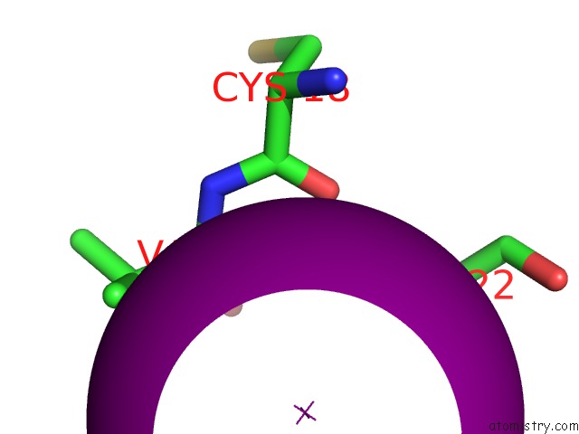
Mono view
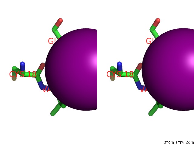
Stereo pair view
Mono view
Stereo pair view
A full contact list of Iodine with other atoms in the I binding
site number 1 of RING1B-BMI1 E3 Catalytic Domain Structure within 5.0Å range:
|
Iodine binding site 2 out of 2 in 2ckl
Go back to
Iodine binding site 2 out
of 2 in the RING1B-BMI1 E3 Catalytic Domain Structure

Mono view

Stereo pair view
Mono view
Stereo pair view
A full contact list of Iodine with other atoms in the I binding
site number 2 of RING1B-BMI1 E3 Catalytic Domain Structure within 5.0Å range:
|
Reference:
G.Buchwald,
P.Van Der Stoop,
O.Weichenrieder,
A.Perrakis,
M.Van Lohuizen,
T.K.Sixma.
Structure and E3-Ligase Activity of the Ring-Ring Complex of Polycomb Proteins BMI1 and RING1B. Embo J. V. 25 2465 2006.
ISSN: ISSN 0261-4189
PubMed: 16710298
DOI: 10.1038/SJ.EMBOJ.7601144
Page generated: Sun Aug 11 13:39:21 2024
ISSN: ISSN 0261-4189
PubMed: 16710298
DOI: 10.1038/SJ.EMBOJ.7601144
Last articles
Zn in 9J0NZn in 9J0O
Zn in 9J0P
Zn in 9FJX
Zn in 9EKB
Zn in 9C0F
Zn in 9CAH
Zn in 9CH0
Zn in 9CH3
Zn in 9CH1