Iodine »
PDB 2axe-2fwh »
2d97 »
Iodine in PDB 2d97: Structure of Vil-Xylanase
Enzymatic activity of Structure of Vil-Xylanase
All present enzymatic activity of Structure of Vil-Xylanase:
3.2.1.8;
3.2.1.8;
Protein crystallography data
The structure of Structure of Vil-Xylanase, PDB code: 2d97
was solved by
H.Miyatake,
T.Hasegawa,
A.Yamano,
with X-Ray Crystallography technique. A brief refinement statistics is given in the table below:
| Resolution Low / High (Å) | 18.98 / 2.01 |
| Space group | P 1 21 1 |
| Cell size a, b, c (Å), α, β, γ (°) | 40.347, 38.585, 57.162, 90.00, 110.31, 90.00 |
| R / Rfree (%) | 19.8 / 22.8 |
Iodine Binding Sites:
The binding sites of Iodine atom in the Structure of Vil-Xylanase
(pdb code 2d97). This binding sites where shown within
5.0 Angstroms radius around Iodine atom.
In total 5 binding sites of Iodine where determined in the Structure of Vil-Xylanase, PDB code: 2d97:
Jump to Iodine binding site number: 1; 2; 3; 4; 5;
In total 5 binding sites of Iodine where determined in the Structure of Vil-Xylanase, PDB code: 2d97:
Jump to Iodine binding site number: 1; 2; 3; 4; 5;
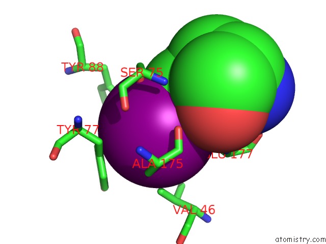
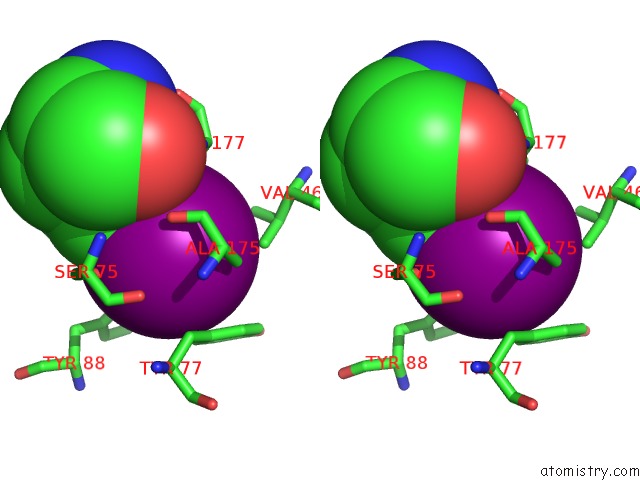
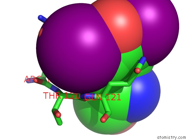
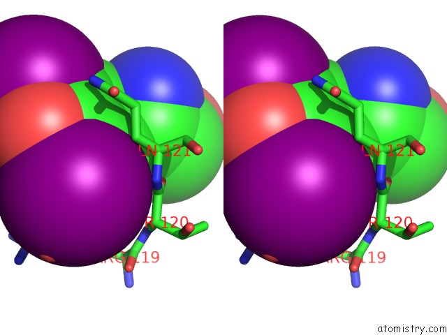
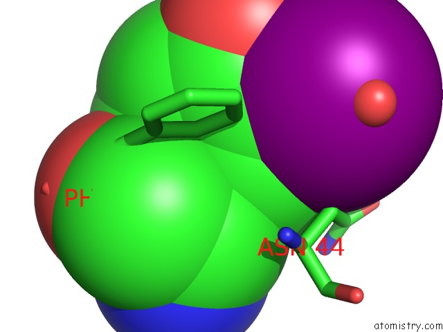
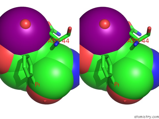
Iodine binding site 1 out of 5 in 2d97
Go back to
Iodine binding site 1 out
of 5 in the Structure of Vil-Xylanase
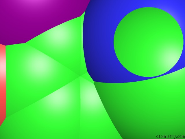
Mono view
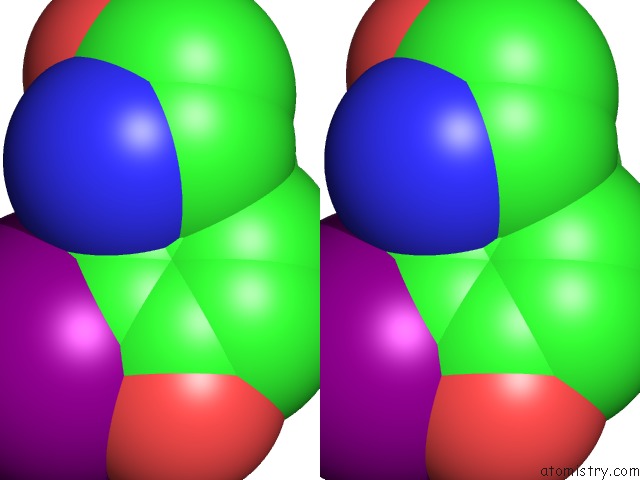
Stereo pair view
Mono view
Stereo pair view
A full contact list of Iodine with other atoms in the I binding
site number 1 of Structure of Vil-Xylanase within 5.0Å range:
|
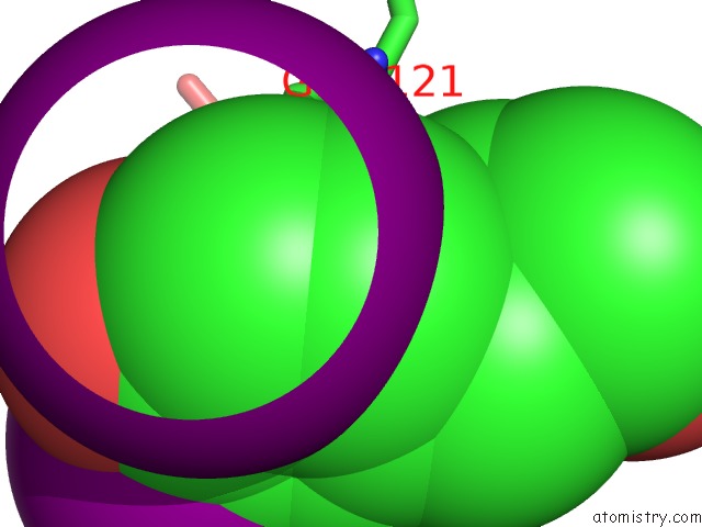
Iodine binding site 2 out of 5 in 2d97
Go back to
Iodine binding site 2 out
of 5 in the Structure of Vil-Xylanase
Mono view
Stereo pair view
Mono view
Stereo pair view
A full contact list of Iodine with other atoms in the I binding
site number 2 of Structure of Vil-Xylanase within 5.0Å range:
|
Iodine binding site 3 out of 5 in 2d97
Go back to
Iodine binding site 3 out
of 5 in the Structure of Vil-Xylanase
Mono view
Stereo pair view
Mono view
Stereo pair view
A full contact list of Iodine with other atoms in the I binding
site number 3 of Structure of Vil-Xylanase within 5.0Å range:
|
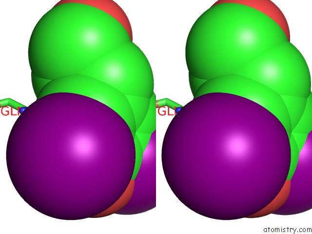
Iodine binding site 4 out of 5 in 2d97
Go back to
Iodine binding site 4 out
of 5 in the Structure of Vil-Xylanase
Mono view
Stereo pair view
Mono view
Stereo pair view
A full contact list of Iodine with other atoms in the I binding
site number 4 of Structure of Vil-Xylanase within 5.0Å range:
|
Iodine binding site 5 out of 5 in 2d97
Go back to
Iodine binding site 5 out
of 5 in the Structure of Vil-Xylanase
Mono view
Stereo pair view
Mono view
Stereo pair view
A full contact list of Iodine with other atoms in the I binding
site number 5 of Structure of Vil-Xylanase within 5.0Å range:
|
Reference:
H.Miyatake,
T.Hasegawa,
A.Yamano.
New Methods to Prepare Iodinated Derivatives By Vaporizing Iodine Labelling (Vil) and Hydrogen Peroxide Vil (Hyper-Vil) Acta Crystallogr.,Sect.D V. 62 280 2006.
ISSN: ISSN 0907-4449
PubMed: 16510975
DOI: 10.1107/S0907444905041909
Page generated: Sun Aug 11 13:43:28 2024
ISSN: ISSN 0907-4449
PubMed: 16510975
DOI: 10.1107/S0907444905041909
Last articles
Zn in 9J0NZn in 9J0O
Zn in 9J0P
Zn in 9FJX
Zn in 9EKB
Zn in 9C0F
Zn in 9CAH
Zn in 9CH0
Zn in 9CH3
Zn in 9CH1