Iodine »
PDB 2axe-2fwh »
2dfc »
Iodine in PDB 2dfc: Xylanase II From Tricoderma Reesei at 293K
Enzymatic activity of Xylanase II From Tricoderma Reesei at 293K
All present enzymatic activity of Xylanase II From Tricoderma Reesei at 293K:
3.2.1.8;
3.2.1.8;
Protein crystallography data
The structure of Xylanase II From Tricoderma Reesei at 293K, PDB code: 2dfc
was solved by
K.Harata,
T.Akiba,
with X-Ray Crystallography technique. A brief refinement statistics is given in the table below:
| Resolution Low / High (Å) | 20.00 / 1.19 |
| Space group | P 21 21 21 |
| Cell size a, b, c (Å), α, β, γ (°) | 49.550, 60.030, 70.500, 90.00, 90.00, 90.00 |
| R / Rfree (%) | 10.6 / 12.2 |
Iodine Binding Sites:
The binding sites of Iodine atom in the Xylanase II From Tricoderma Reesei at 293K
(pdb code 2dfc). This binding sites where shown within
5.0 Angstroms radius around Iodine atom.
In total only one binding site of Iodine was determined in the Xylanase II From Tricoderma Reesei at 293K, PDB code: 2dfc:
In total only one binding site of Iodine was determined in the Xylanase II From Tricoderma Reesei at 293K, PDB code: 2dfc:
Iodine binding site 1 out of 1 in 2dfc
Go back to
Iodine binding site 1 out
of 1 in the Xylanase II From Tricoderma Reesei at 293K
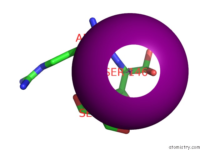
Mono view
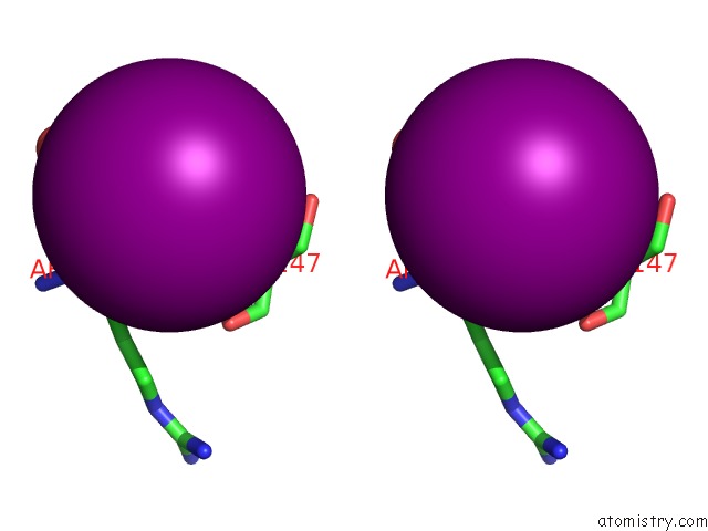
Stereo pair view
Mono view
Stereo pair view
A full contact list of Iodine with other atoms in the I binding
site number 1 of Xylanase II From Tricoderma Reesei at 293K within 5.0Å range:
|
Reference:
N.Watanabe,
T.Akiba,
R.Kanai,
K.Harata.
Structure of An Orthorhombic Form of Xylanase II From Trichoderma Reesei and Analysis of Thermal Displacement. Acta Crystallogr.,Sect.D V. 62 784 2006.
ISSN: ISSN 0907-4449
PubMed: 16790934
DOI: 10.1107/S0907444906017379
Page generated: Sun Aug 11 13:45:05 2024
ISSN: ISSN 0907-4449
PubMed: 16790934
DOI: 10.1107/S0907444906017379
Last articles
Zn in 9MJ5Zn in 9HNW
Zn in 9G0L
Zn in 9FNE
Zn in 9DZN
Zn in 9E0I
Zn in 9D32
Zn in 9DAK
Zn in 8ZXC
Zn in 8ZUF