Iodine »
PDB 2axe-2fwh »
2dgm »
Iodine in PDB 2dgm: Crystal Structure of Escherichia Coli Gadb in Complex with Iodide
Enzymatic activity of Crystal Structure of Escherichia Coli Gadb in Complex with Iodide
All present enzymatic activity of Crystal Structure of Escherichia Coli Gadb in Complex with Iodide:
4.1.1.15;
4.1.1.15;
Protein crystallography data
The structure of Crystal Structure of Escherichia Coli Gadb in Complex with Iodide, PDB code: 2dgm
was solved by
M.G.Gruetter,
G.Capitani,
H.Gut,
with X-Ray Crystallography technique. A brief refinement statistics is given in the table below:
| Resolution Low / High (Å) | 40.00 / 1.95 |
| Space group | P 1 |
| Cell size a, b, c (Å), α, β, γ (°) | 91.336, 91.804, 93.914, 76.65, 76.94, 78.04 |
| R / Rfree (%) | 22.7 / 26.7 |
Iodine Binding Sites:
Pages:
>>> Page 1 <<< Page 2, Binding sites: 11 - 20; Page 3, Binding sites: 21 - 30; Page 4, Binding sites: 31 - 32;Binding sites:
The binding sites of Iodine atom in the Crystal Structure of Escherichia Coli Gadb in Complex with Iodide (pdb code 2dgm). This binding sites where shown within 5.0 Angstroms radius around Iodine atom.In total 32 binding sites of Iodine where determined in the Crystal Structure of Escherichia Coli Gadb in Complex with Iodide, PDB code: 2dgm:
Jump to Iodine binding site number: 1; 2; 3; 4; 5; 6; 7; 8; 9; 10;
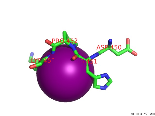
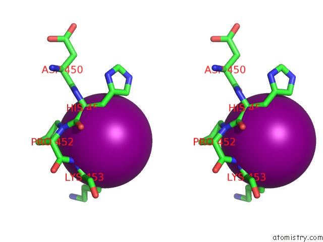
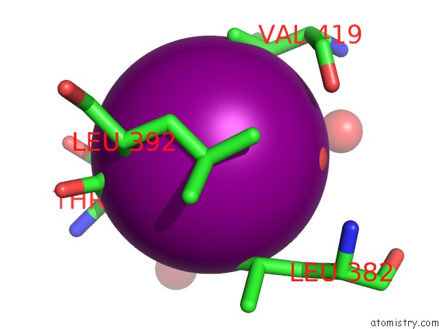
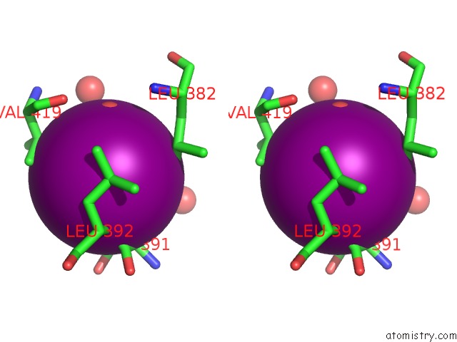
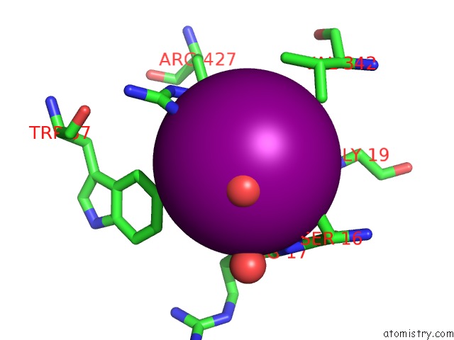
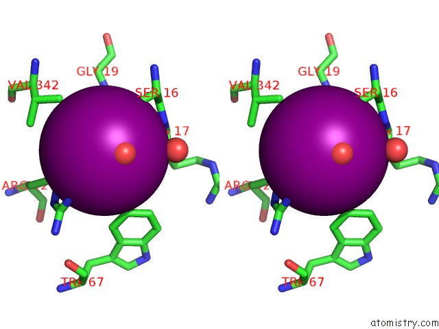
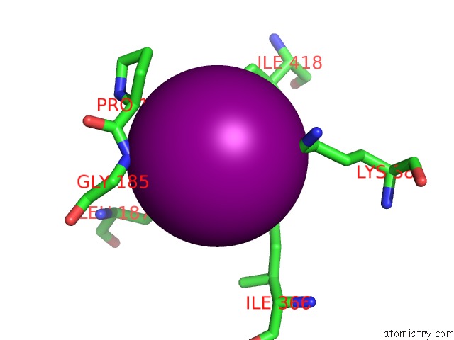
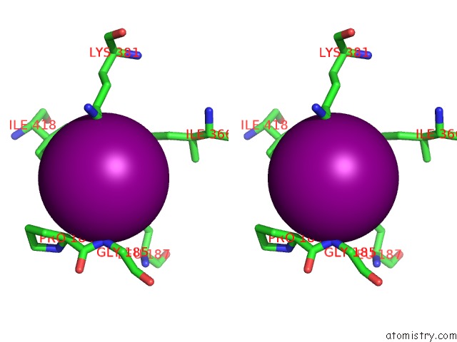
Iodine binding site 1 out of 32 in 2dgm
Go back to
Iodine binding site 1 out
of 32 in the Crystal Structure of Escherichia Coli Gadb in Complex with Iodide
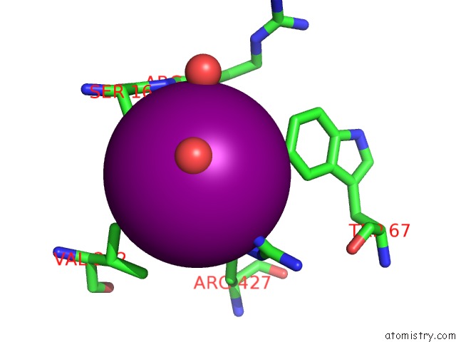
Mono view
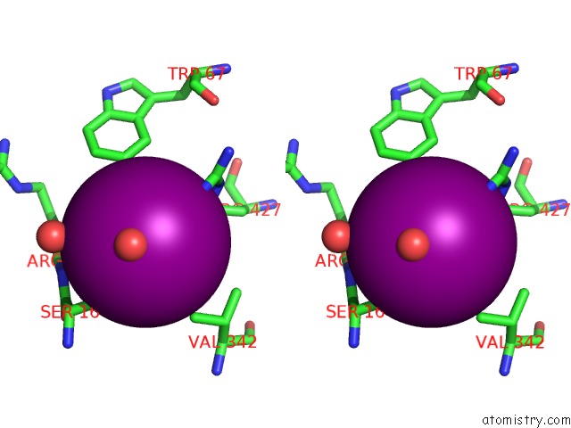
Stereo pair view
Mono view
Stereo pair view
A full contact list of Iodine with other atoms in the I binding
site number 1 of Crystal Structure of Escherichia Coli Gadb in Complex with Iodide within 5.0Å range:
|
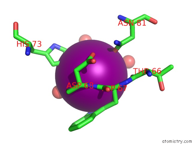
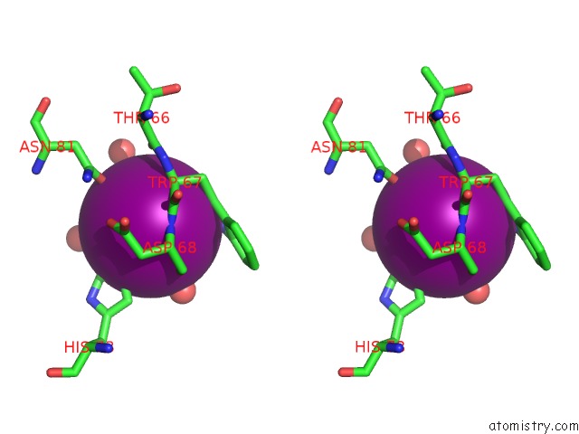
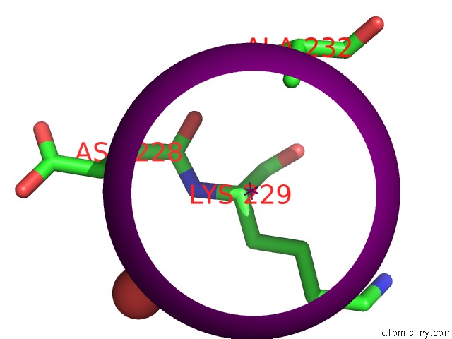
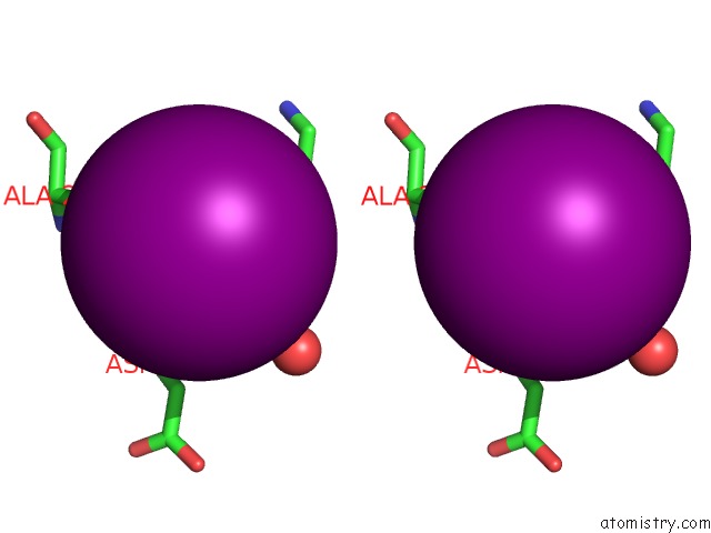
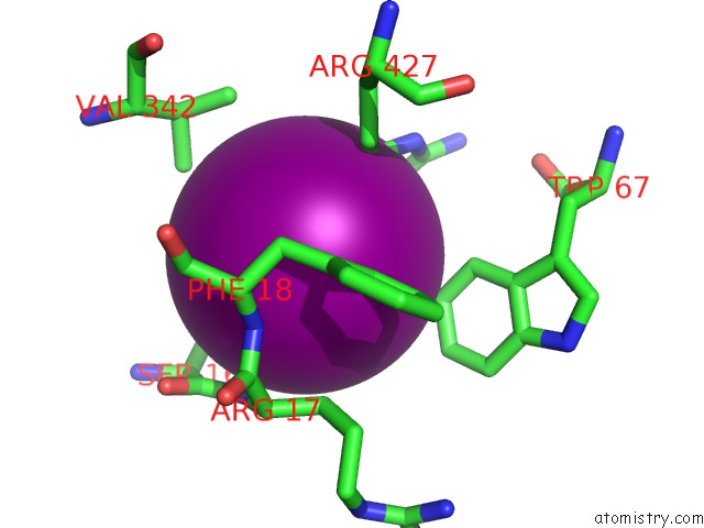
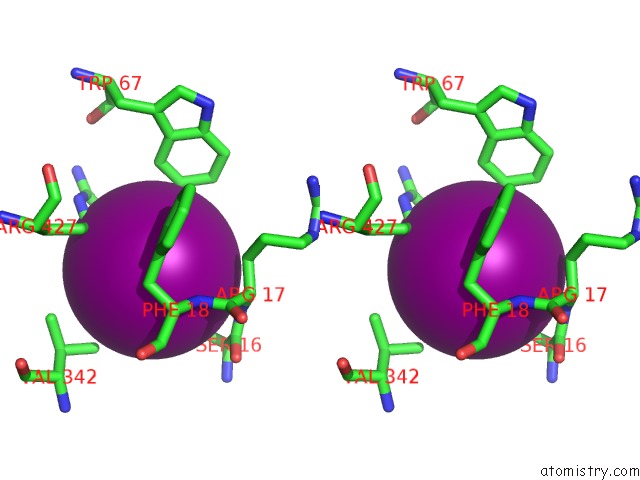
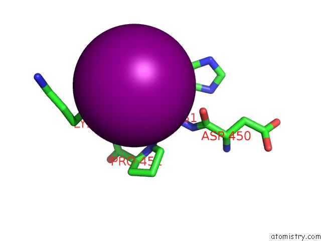
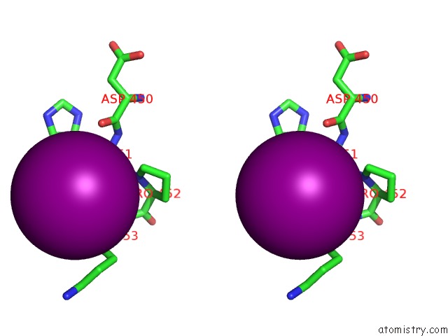
Iodine binding site 2 out of 32 in 2dgm
Go back to
Iodine binding site 2 out
of 32 in the Crystal Structure of Escherichia Coli Gadb in Complex with Iodide
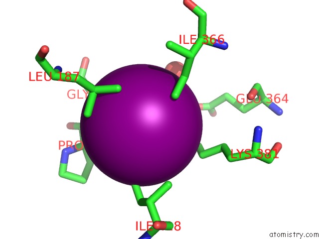
Mono view
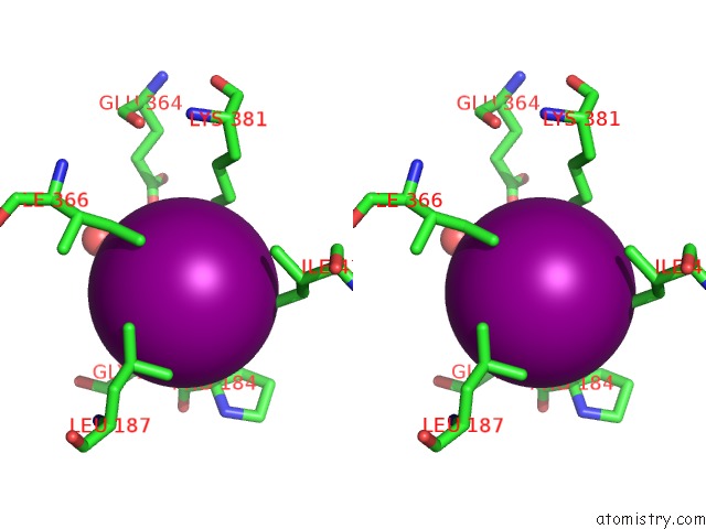
Stereo pair view
Mono view
Stereo pair view
A full contact list of Iodine with other atoms in the I binding
site number 2 of Crystal Structure of Escherichia Coli Gadb in Complex with Iodide within 5.0Å range:
|
Iodine binding site 3 out of 32 in 2dgm
Go back to
Iodine binding site 3 out
of 32 in the Crystal Structure of Escherichia Coli Gadb in Complex with Iodide
Mono view
Stereo pair view
Mono view
Stereo pair view
A full contact list of Iodine with other atoms in the I binding
site number 3 of Crystal Structure of Escherichia Coli Gadb in Complex with Iodide within 5.0Å range:
|
Iodine binding site 4 out of 32 in 2dgm
Go back to
Iodine binding site 4 out
of 32 in the Crystal Structure of Escherichia Coli Gadb in Complex with Iodide
Mono view
Stereo pair view
Mono view
Stereo pair view
A full contact list of Iodine with other atoms in the I binding
site number 4 of Crystal Structure of Escherichia Coli Gadb in Complex with Iodide within 5.0Å range:
|
Iodine binding site 5 out of 32 in 2dgm
Go back to
Iodine binding site 5 out
of 32 in the Crystal Structure of Escherichia Coli Gadb in Complex with Iodide
Mono view
Stereo pair view
Mono view
Stereo pair view
A full contact list of Iodine with other atoms in the I binding
site number 5 of Crystal Structure of Escherichia Coli Gadb in Complex with Iodide within 5.0Å range:
|
Iodine binding site 6 out of 32 in 2dgm
Go back to
Iodine binding site 6 out
of 32 in the Crystal Structure of Escherichia Coli Gadb in Complex with Iodide
Mono view
Stereo pair view
Mono view
Stereo pair view
A full contact list of Iodine with other atoms in the I binding
site number 6 of Crystal Structure of Escherichia Coli Gadb in Complex with Iodide within 5.0Å range:
|
Iodine binding site 7 out of 32 in 2dgm
Go back to
Iodine binding site 7 out
of 32 in the Crystal Structure of Escherichia Coli Gadb in Complex with Iodide
Mono view
Stereo pair view
Mono view
Stereo pair view
A full contact list of Iodine with other atoms in the I binding
site number 7 of Crystal Structure of Escherichia Coli Gadb in Complex with Iodide within 5.0Å range:
|
Iodine binding site 8 out of 32 in 2dgm
Go back to
Iodine binding site 8 out
of 32 in the Crystal Structure of Escherichia Coli Gadb in Complex with Iodide
Mono view
Stereo pair view
Mono view
Stereo pair view
A full contact list of Iodine with other atoms in the I binding
site number 8 of Crystal Structure of Escherichia Coli Gadb in Complex with Iodide within 5.0Å range:
|
Iodine binding site 9 out of 32 in 2dgm
Go back to
Iodine binding site 9 out
of 32 in the Crystal Structure of Escherichia Coli Gadb in Complex with Iodide
Mono view
Stereo pair view
Mono view
Stereo pair view
A full contact list of Iodine with other atoms in the I binding
site number 9 of Crystal Structure of Escherichia Coli Gadb in Complex with Iodide within 5.0Å range:
|
Iodine binding site 10 out of 32 in 2dgm
Go back to
Iodine binding site 10 out
of 32 in the Crystal Structure of Escherichia Coli Gadb in Complex with Iodide
Mono view
Stereo pair view
Mono view
Stereo pair view
A full contact list of Iodine with other atoms in the I binding
site number 10 of Crystal Structure of Escherichia Coli Gadb in Complex with Iodide within 5.0Å range:
|
Reference:
H.Gut,
E.Pennacchietti,
R.A.John,
F.Bossa,
G.Capitani,
D.De Biase,
M.G.Gruetter.
Escherichia Coli Acid Resistance: pH-Sensing, Activation By Chloride and Autoinhibition in Gadb Embo J. V. 25 2643 2006.
ISSN: ISSN 0261-4189
PubMed: 16675957
DOI: 10.1038/SJ.EMBOJ.7601107
Page generated: Sun Aug 11 13:45:19 2024
ISSN: ISSN 0261-4189
PubMed: 16675957
DOI: 10.1038/SJ.EMBOJ.7601107
Last articles
F in 7KUOF in 7KUN
F in 7KRO
F in 7KUI
F in 7KUE
F in 7KU7
F in 7KRN
F in 7KSI
F in 7KSJ
F in 7KQJ