Iodine »
PDB 2axe-2fwh »
2fwh »
Iodine in PDB 2fwh: Atomic Resolution Crystal Structure of the C-Terminal Domain of the Electron Transfer Catalyst Dsbd (Reduced Form at PH7)
Enzymatic activity of Atomic Resolution Crystal Structure of the C-Terminal Domain of the Electron Transfer Catalyst Dsbd (Reduced Form at PH7)
All present enzymatic activity of Atomic Resolution Crystal Structure of the C-Terminal Domain of the Electron Transfer Catalyst Dsbd (Reduced Form at PH7):
1.8.1.8;
1.8.1.8;
Protein crystallography data
The structure of Atomic Resolution Crystal Structure of the C-Terminal Domain of the Electron Transfer Catalyst Dsbd (Reduced Form at PH7), PDB code: 2fwh
was solved by
C.U.Stirnimann,
A.Rozhkova,
U.Grauschopf,
R.A.Boeckmann,
R.Glockshuber,
G.Capitani,
M.G.Gruetter,
with X-Ray Crystallography technique. A brief refinement statistics is given in the table below:
| Resolution Low / High (Å) | 15.00 / 0.99 |
| Space group | P 21 21 21 |
| Cell size a, b, c (Å), α, β, γ (°) | 30.289, 46.072, 74.070, 90.00, 90.00, 90.00 |
| R / Rfree (%) | n/a / 14.6 |
Iodine Binding Sites:
The binding sites of Iodine atom in the Atomic Resolution Crystal Structure of the C-Terminal Domain of the Electron Transfer Catalyst Dsbd (Reduced Form at PH7)
(pdb code 2fwh). This binding sites where shown within
5.0 Angstroms radius around Iodine atom.
In total 3 binding sites of Iodine where determined in the Atomic Resolution Crystal Structure of the C-Terminal Domain of the Electron Transfer Catalyst Dsbd (Reduced Form at PH7), PDB code: 2fwh:
Jump to Iodine binding site number: 1; 2; 3;
In total 3 binding sites of Iodine where determined in the Atomic Resolution Crystal Structure of the C-Terminal Domain of the Electron Transfer Catalyst Dsbd (Reduced Form at PH7), PDB code: 2fwh:
Jump to Iodine binding site number: 1; 2; 3;
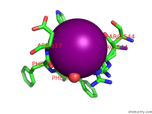
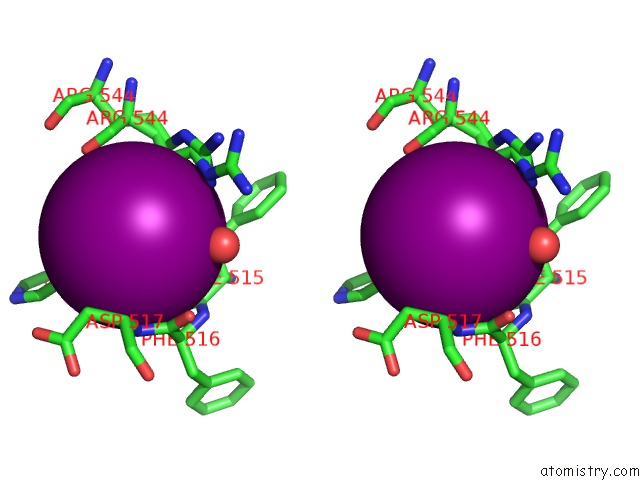
Iodine binding site 1 out of 3 in 2fwh
Go back to
Iodine binding site 1 out
of 3 in the Atomic Resolution Crystal Structure of the C-Terminal Domain of the Electron Transfer Catalyst Dsbd (Reduced Form at PH7)
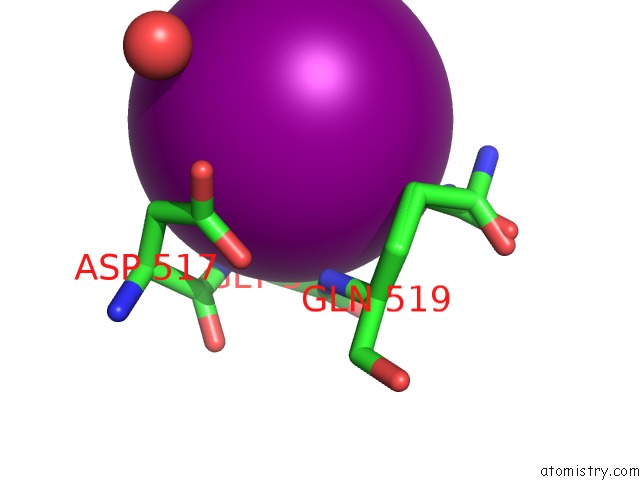
Mono view
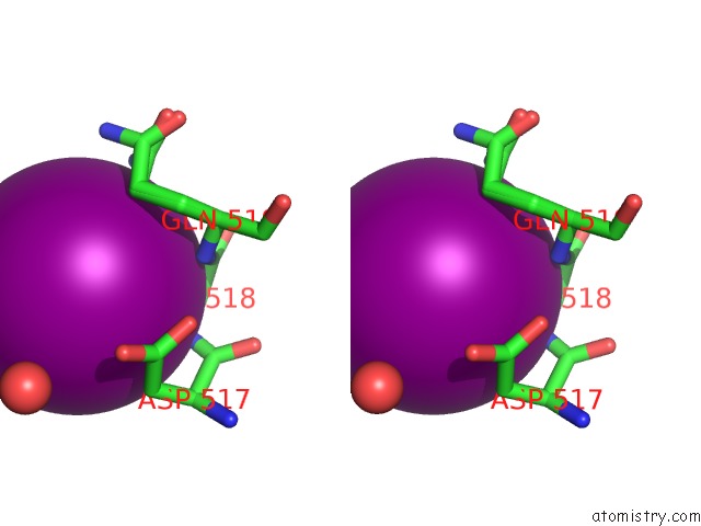
Stereo pair view
Mono view
Stereo pair view
A full contact list of Iodine with other atoms in the I binding
site number 1 of Atomic Resolution Crystal Structure of the C-Terminal Domain of the Electron Transfer Catalyst Dsbd (Reduced Form at PH7) within 5.0Å range:
|
Iodine binding site 2 out of 3 in 2fwh
Go back to
Iodine binding site 2 out
of 3 in the Atomic Resolution Crystal Structure of the C-Terminal Domain of the Electron Transfer Catalyst Dsbd (Reduced Form at PH7)
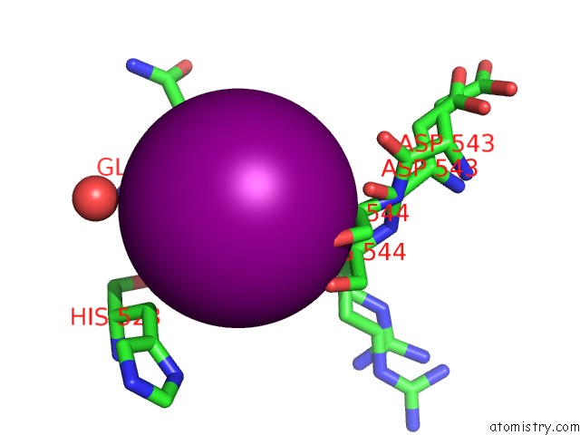
Mono view
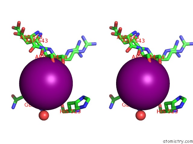
Stereo pair view
Mono view
Stereo pair view
A full contact list of Iodine with other atoms in the I binding
site number 2 of Atomic Resolution Crystal Structure of the C-Terminal Domain of the Electron Transfer Catalyst Dsbd (Reduced Form at PH7) within 5.0Å range:
|
Iodine binding site 3 out of 3 in 2fwh
Go back to
Iodine binding site 3 out
of 3 in the Atomic Resolution Crystal Structure of the C-Terminal Domain of the Electron Transfer Catalyst Dsbd (Reduced Form at PH7)
Mono view
Stereo pair view
Mono view
Stereo pair view
A full contact list of Iodine with other atoms in the I binding
site number 3 of Atomic Resolution Crystal Structure of the C-Terminal Domain of the Electron Transfer Catalyst Dsbd (Reduced Form at PH7) within 5.0Å range:
|
Reference:
C.U.Stirnimann,
A.Rozhkova,
U.Grauschopf,
R.A.Boeckmann,
R.Glockshuber,
G.Capitani,
M.G.Gruetter.
High-Resolution Structures of Escherichia Coli Cdsbd in Different Redox States: A Combined Crystallographic, Biochemical and Computational Study J.Mol.Biol. V. 358 829 2006.
ISSN: ISSN 0022-2836
PubMed: 16545842
DOI: 10.1016/J.JMB.2006.02.030
Page generated: Sun Aug 11 13:49:10 2024
ISSN: ISSN 0022-2836
PubMed: 16545842
DOI: 10.1016/J.JMB.2006.02.030
Last articles
Cl in 2W2DCl in 2W2J
Cl in 2W2E
Cl in 2W3K
Cl in 2W3I
Cl in 2W1Y
Cl in 2W26
Cl in 2W20
Cl in 2W1X
Cl in 2VXA