Iodine »
PDB 2fwz-2noo »
2g1m »
Iodine in PDB 2g1m: Cellular Oxygen Sensing: Crystal Structure of Hypoxia-Inducible Factor Prolyl Hydroxylase (PHD2)
Protein crystallography data
The structure of Cellular Oxygen Sensing: Crystal Structure of Hypoxia-Inducible Factor Prolyl Hydroxylase (PHD2), PDB code: 2g1m
was solved by
M.A.Mcdonough,
C.J.Schofield,
with X-Ray Crystallography technique. A brief refinement statistics is given in the table below:
| Resolution Low / High (Å) | 30.71 / 2.20 |
| Space group | P 63 |
| Cell size a, b, c (Å), α, β, γ (°) | 110.742, 110.742, 39.986, 90.00, 90.00, 120.00 |
| R / Rfree (%) | 21.6 / 28.9 |
Other elements in 2g1m:
The structure of Cellular Oxygen Sensing: Crystal Structure of Hypoxia-Inducible Factor Prolyl Hydroxylase (PHD2) also contains other interesting chemical elements:
| Iron | (Fe) | 1 atom |
Iodine Binding Sites:
The binding sites of Iodine atom in the Cellular Oxygen Sensing: Crystal Structure of Hypoxia-Inducible Factor Prolyl Hydroxylase (PHD2)
(pdb code 2g1m). This binding sites where shown within
5.0 Angstroms radius around Iodine atom.
In total only one binding site of Iodine was determined in the Cellular Oxygen Sensing: Crystal Structure of Hypoxia-Inducible Factor Prolyl Hydroxylase (PHD2), PDB code: 2g1m:
In total only one binding site of Iodine was determined in the Cellular Oxygen Sensing: Crystal Structure of Hypoxia-Inducible Factor Prolyl Hydroxylase (PHD2), PDB code: 2g1m:
Iodine binding site 1 out of 1 in 2g1m
Go back to
Iodine binding site 1 out
of 1 in the Cellular Oxygen Sensing: Crystal Structure of Hypoxia-Inducible Factor Prolyl Hydroxylase (PHD2)
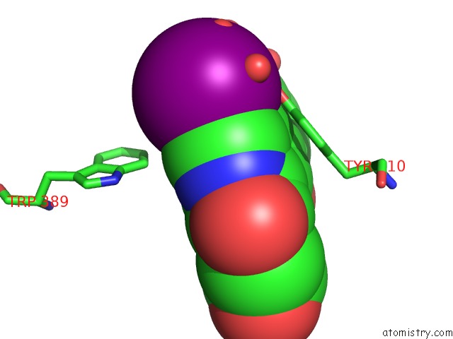
Mono view
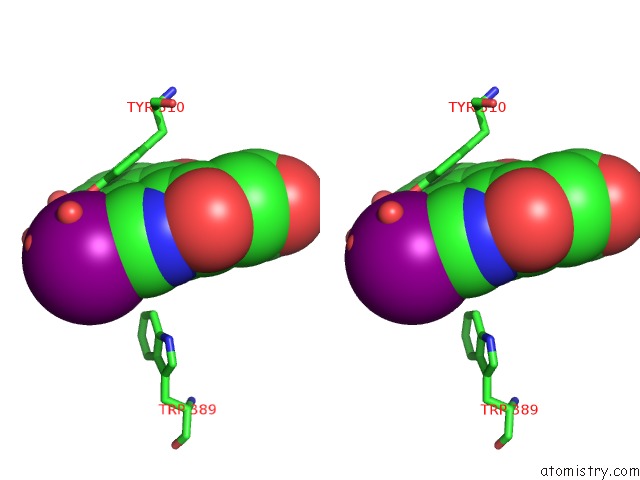
Stereo pair view
Mono view
Stereo pair view
A full contact list of Iodine with other atoms in the I binding
site number 1 of Cellular Oxygen Sensing: Crystal Structure of Hypoxia-Inducible Factor Prolyl Hydroxylase (PHD2) within 5.0Å range:
|
Reference:
M.A.Mcdonough,
V.Li,
E.Flashman,
R.Chowdhury,
C.Mohr,
B.M.Lienard,
J.Zondlo,
N.J.Oldham,
I.J.Clifton,
J.Lewis,
L.A.Mcneill,
R.J.Kurzeja,
K.S.Hewitson,
E.Yang,
S.Jordan,
R.S.Syed,
C.J.Schofield.
Cellular Oxygen Sensing: Crystal Structure of Hypoxia-Inducible Factor Prolyl Hydroxylase (PHD2). Proc.Natl.Acad.Sci.Usa V. 103 9814 2006.
ISSN: ISSN 0027-8424
PubMed: 16782814
DOI: 10.1073/PNAS.0601283103
Page generated: Sun Aug 11 13:52:11 2024
ISSN: ISSN 0027-8424
PubMed: 16782814
DOI: 10.1073/PNAS.0601283103
Last articles
Zn in 9J0NZn in 9J0O
Zn in 9J0P
Zn in 9FJX
Zn in 9EKB
Zn in 9C0F
Zn in 9CAH
Zn in 9CH0
Zn in 9CH3
Zn in 9CH1