Iodine »
PDB 2fwz-2noo »
2h77 »
Iodine in PDB 2h77: Crystal Structure of Human Tr Alpha Bound T3 in Monoclinic Space Group
Protein crystallography data
The structure of Crystal Structure of Human Tr Alpha Bound T3 in Monoclinic Space Group, PDB code: 2h77
was solved by
A.S.Nascimento,
S.M.G.Dias,
F.M.Nunes,
R.Aparicio,
L.Bleicher,
A.L.B.Ambrosio,
A.C.M.Figueira,
M.A.M.Santos,
M.O.Neto,
H.Fischer,
H.F.M.Togashi,
A.F.Craievich,
R.C.Garrat,
J.D.Baxter,
P.Webb,
I.Polikarpov,
with X-Ray Crystallography technique. A brief refinement statistics is given in the table below:
| Resolution Low / High (Å) | 32.22 / 2.33 |
| Space group | C 1 2 1 |
| Cell size a, b, c (Å), α, β, γ (°) | 117.569, 80.626, 62.614, 90.00, 121.09, 90.00 |
| R / Rfree (%) | 18.7 / 23.8 |
Other elements in 2h77:
The structure of Crystal Structure of Human Tr Alpha Bound T3 in Monoclinic Space Group also contains other interesting chemical elements:
| Arsenic | (As) | 4 atoms |
Iodine Binding Sites:
The binding sites of Iodine atom in the Crystal Structure of Human Tr Alpha Bound T3 in Monoclinic Space Group
(pdb code 2h77). This binding sites where shown within
5.0 Angstroms radius around Iodine atom.
In total 3 binding sites of Iodine where determined in the Crystal Structure of Human Tr Alpha Bound T3 in Monoclinic Space Group, PDB code: 2h77:
Jump to Iodine binding site number: 1; 2; 3;
In total 3 binding sites of Iodine where determined in the Crystal Structure of Human Tr Alpha Bound T3 in Monoclinic Space Group, PDB code: 2h77:
Jump to Iodine binding site number: 1; 2; 3;
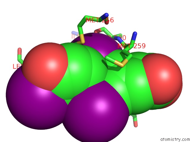
Iodine binding site 1 out of 3 in 2h77
Go back to
Iodine binding site 1 out
of 3 in the Crystal Structure of Human Tr Alpha Bound T3 in Monoclinic Space Group
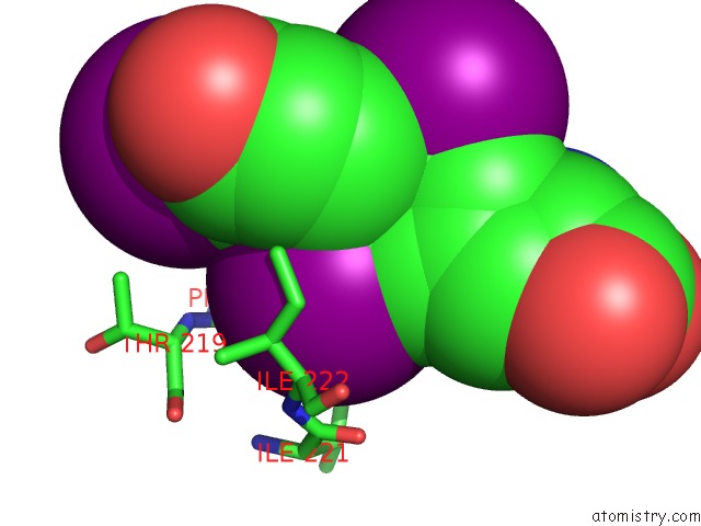
Mono view
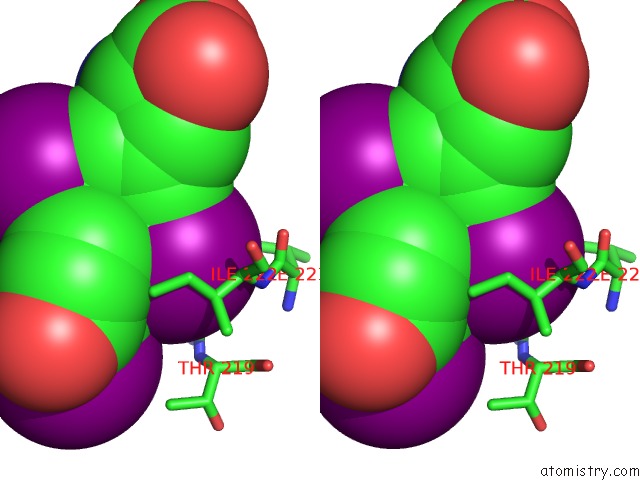
Stereo pair view
Mono view
Stereo pair view
A full contact list of Iodine with other atoms in the I binding
site number 1 of Crystal Structure of Human Tr Alpha Bound T3 in Monoclinic Space Group within 5.0Å range:
|
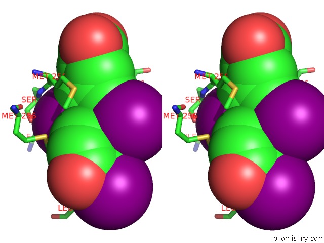
Iodine binding site 2 out of 3 in 2h77
Go back to
Iodine binding site 2 out
of 3 in the Crystal Structure of Human Tr Alpha Bound T3 in Monoclinic Space Group
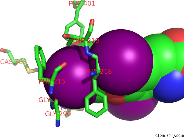
Mono view
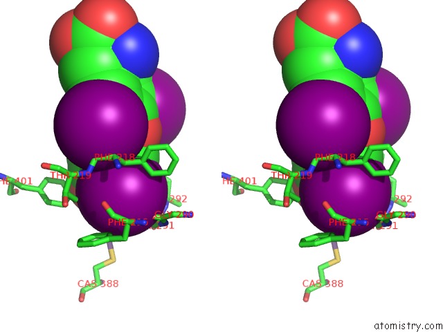
Stereo pair view
Mono view
Stereo pair view
A full contact list of Iodine with other atoms in the I binding
site number 2 of Crystal Structure of Human Tr Alpha Bound T3 in Monoclinic Space Group within 5.0Å range:
|
Iodine binding site 3 out of 3 in 2h77
Go back to
Iodine binding site 3 out
of 3 in the Crystal Structure of Human Tr Alpha Bound T3 in Monoclinic Space Group
Mono view
Stereo pair view
Mono view
Stereo pair view
A full contact list of Iodine with other atoms in the I binding
site number 3 of Crystal Structure of Human Tr Alpha Bound T3 in Monoclinic Space Group within 5.0Å range:
|
Reference:
A.S.Nascimento,
S.M.G.Dias,
F.M.Nunes,
R.Aparicio,
A.L.B.Ambrosio,
L.Bleicher,
A.C.M.Figueira,
M.A.M.Santos,
M.O.Neto,
H.Fischer,
M.Togashi,
A.F.Craievich,
R.C.Garratt,
J.D.Baxter,
P.Webb,
I.Polikarpov.
Structural Rearrangements in the Thyroid Hormone Receptor Hinge Domain and Their Putative Role in the Receptor Function. J.Mol.Biol. V. 360 586 2006.
ISSN: ISSN 0022-2836
PubMed: 16781732
DOI: 10.1016/J.JMB.2006.05.008
Page generated: Sun Aug 11 13:54:07 2024
ISSN: ISSN 0022-2836
PubMed: 16781732
DOI: 10.1016/J.JMB.2006.05.008
Last articles
Zn in 9J0NZn in 9J0O
Zn in 9J0P
Zn in 9FJX
Zn in 9EKB
Zn in 9C0F
Zn in 9CAH
Zn in 9CH0
Zn in 9CH3
Zn in 9CH1