Iodine »
PDB 2fwz-2noo »
2noo »
Iodine in PDB 2noo: Crystal Structure of Mutant Nika
Enzymatic activity of Crystal Structure of Mutant Nika
All present enzymatic activity of Crystal Structure of Mutant Nika:
3.6.3.24;
3.6.3.24;
Protein crystallography data
The structure of Crystal Structure of Mutant Nika, PDB code: 2noo
was solved by
C.Addy,
M.Ohara,
F.Kawai,
A.Kidera,
M.Ikeguchi,
S.Fuchigami,
M.Osawa,
I.Shimada,
S.Y.Park,
J.R.H.Tame,
J.G.Heddle,
with X-Ray Crystallography technique. A brief refinement statistics is given in the table below:
| Resolution Low / High (Å) | 19.82 / 1.65 |
| Space group | P 21 21 21 |
| Cell size a, b, c (Å), α, β, γ (°) | 43.470, 92.248, 116.383, 90.00, 90.00, 90.00 |
| R / Rfree (%) | 17.7 / 21 |
Other elements in 2noo:
The structure of Crystal Structure of Mutant Nika also contains other interesting chemical elements:
| Nickel | (Ni) | 1 atom |
Iodine Binding Sites:
The binding sites of Iodine atom in the Crystal Structure of Mutant Nika
(pdb code 2noo). This binding sites where shown within
5.0 Angstroms radius around Iodine atom.
In total 6 binding sites of Iodine where determined in the Crystal Structure of Mutant Nika, PDB code: 2noo:
Jump to Iodine binding site number: 1; 2; 3; 4; 5; 6;
In total 6 binding sites of Iodine where determined in the Crystal Structure of Mutant Nika, PDB code: 2noo:
Jump to Iodine binding site number: 1; 2; 3; 4; 5; 6;
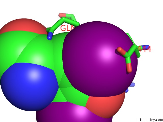
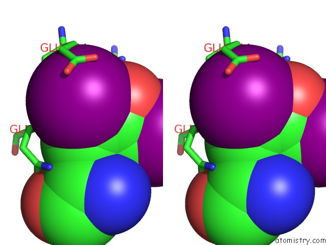
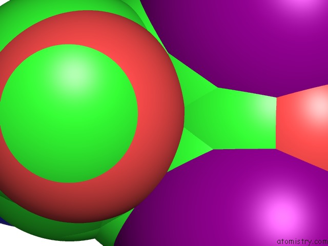
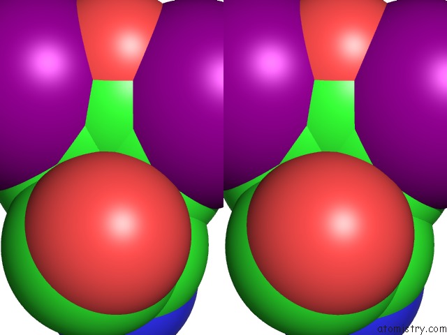
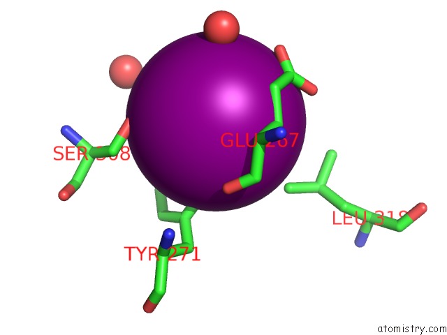
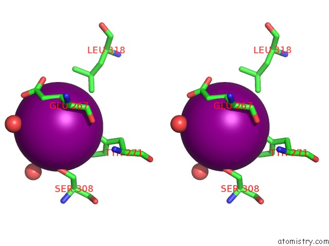
Iodine binding site 1 out of 6 in 2noo
Go back to
Iodine binding site 1 out
of 6 in the Crystal Structure of Mutant Nika
Mono view
Stereo pair view
Mono view
Stereo pair view
A full contact list of Iodine with other atoms in the I binding
site number 1 of Crystal Structure of Mutant Nika within 5.0Å range:
|
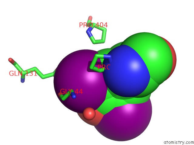
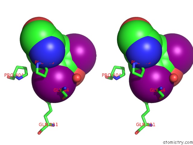
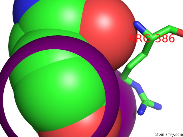
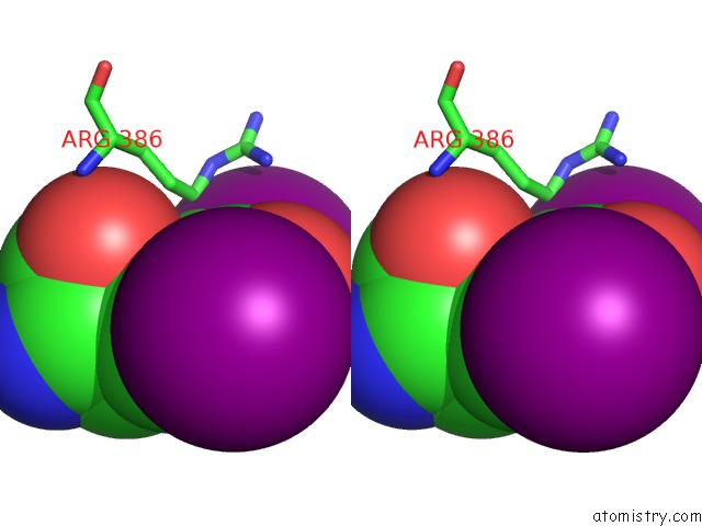
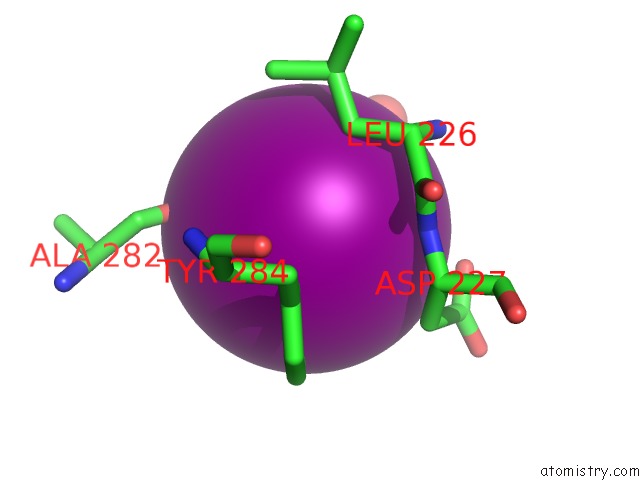
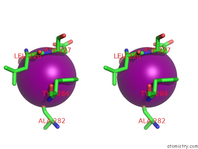
Iodine binding site 2 out of 6 in 2noo
Go back to
Iodine binding site 2 out
of 6 in the Crystal Structure of Mutant Nika
Mono view
Stereo pair view
Mono view
Stereo pair view
A full contact list of Iodine with other atoms in the I binding
site number 2 of Crystal Structure of Mutant Nika within 5.0Å range:
|
Iodine binding site 3 out of 6 in 2noo
Go back to
Iodine binding site 3 out
of 6 in the Crystal Structure of Mutant Nika
Mono view
Stereo pair view
Mono view
Stereo pair view
A full contact list of Iodine with other atoms in the I binding
site number 3 of Crystal Structure of Mutant Nika within 5.0Å range:
|
Iodine binding site 4 out of 6 in 2noo
Go back to
Iodine binding site 4 out
of 6 in the Crystal Structure of Mutant Nika
Mono view
Stereo pair view
Mono view
Stereo pair view
A full contact list of Iodine with other atoms in the I binding
site number 4 of Crystal Structure of Mutant Nika within 5.0Å range:
|
Iodine binding site 5 out of 6 in 2noo
Go back to
Iodine binding site 5 out
of 6 in the Crystal Structure of Mutant Nika
Mono view
Stereo pair view
Mono view
Stereo pair view
A full contact list of Iodine with other atoms in the I binding
site number 5 of Crystal Structure of Mutant Nika within 5.0Å range:
|
Iodine binding site 6 out of 6 in 2noo
Go back to
Iodine binding site 6 out
of 6 in the Crystal Structure of Mutant Nika
Mono view
Stereo pair view
Mono view
Stereo pair view
A full contact list of Iodine with other atoms in the I binding
site number 6 of Crystal Structure of Mutant Nika within 5.0Å range:
|
Reference:
C.Addy,
M.Ohara,
F.Kawai,
A.Kidera,
M.Ikeguchi,
S.Fuchigami,
M.Osawa,
I.Shimada,
S.Y.Park,
J.R.Tame,
J.G.Heddle.
Nickel Binding to Nika: An Additional Binding Site Reconciles Spectroscopy, Calorimetry and Crystallography. Acta Crystallogr.,Sect.D V. 63 221 2007.
ISSN: ISSN 0907-4449
PubMed: 17242515
DOI: 10.1107/S0907444906048712
Page generated: Sun Aug 11 14:08:06 2024
ISSN: ISSN 0907-4449
PubMed: 17242515
DOI: 10.1107/S0907444906048712
Last articles
Zn in 9J0NZn in 9J0O
Zn in 9J0P
Zn in 9FJX
Zn in 9EKB
Zn in 9C0F
Zn in 9CAH
Zn in 9CH0
Zn in 9CH3
Zn in 9CH1