Iodine »
PDB 2nqx-2qpy »
2of0 »
Iodine in PDB 2of0: X-Ray Crystal Structure of Beta Secretase Complexed with Compound 5
Enzymatic activity of X-Ray Crystal Structure of Beta Secretase Complexed with Compound 5
All present enzymatic activity of X-Ray Crystal Structure of Beta Secretase Complexed with Compound 5:
3.4.23.46;
3.4.23.46;
Protein crystallography data
The structure of X-Ray Crystal Structure of Beta Secretase Complexed with Compound 5, PDB code: 2of0
was solved by
S.Patel,
with X-Ray Crystallography technique. A brief refinement statistics is given in the table below:
| Resolution Low / High (Å) | 38.19 / 2.25 |
| Space group | P 61 2 2 |
| Cell size a, b, c (Å), α, β, γ (°) | 102.412, 102.412, 169.237, 90.00, 90.00, 120.00 |
| R / Rfree (%) | 22 / 28 |
Iodine Binding Sites:
The binding sites of Iodine atom in the X-Ray Crystal Structure of Beta Secretase Complexed with Compound 5
(pdb code 2of0). This binding sites where shown within
5.0 Angstroms radius around Iodine atom.
In total 3 binding sites of Iodine where determined in the X-Ray Crystal Structure of Beta Secretase Complexed with Compound 5, PDB code: 2of0:
Jump to Iodine binding site number: 1; 2; 3;
In total 3 binding sites of Iodine where determined in the X-Ray Crystal Structure of Beta Secretase Complexed with Compound 5, PDB code: 2of0:
Jump to Iodine binding site number: 1; 2; 3;
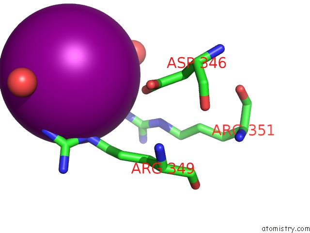
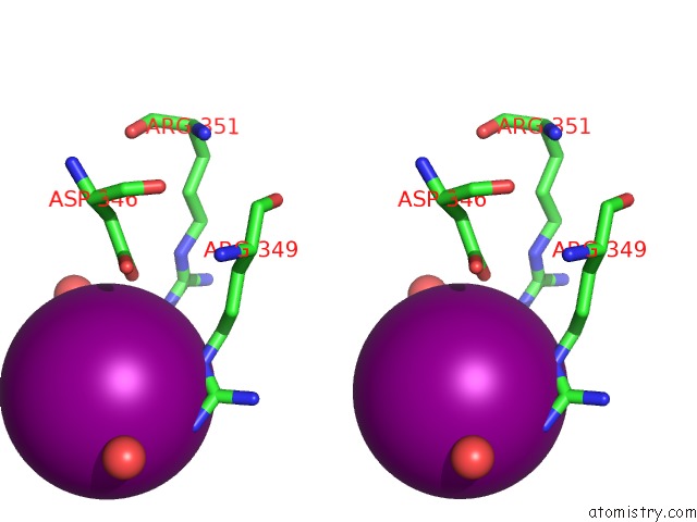
Iodine binding site 1 out of 3 in 2of0
Go back to
Iodine binding site 1 out
of 3 in the X-Ray Crystal Structure of Beta Secretase Complexed with Compound 5
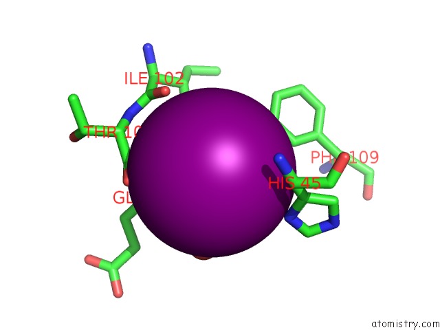
Mono view
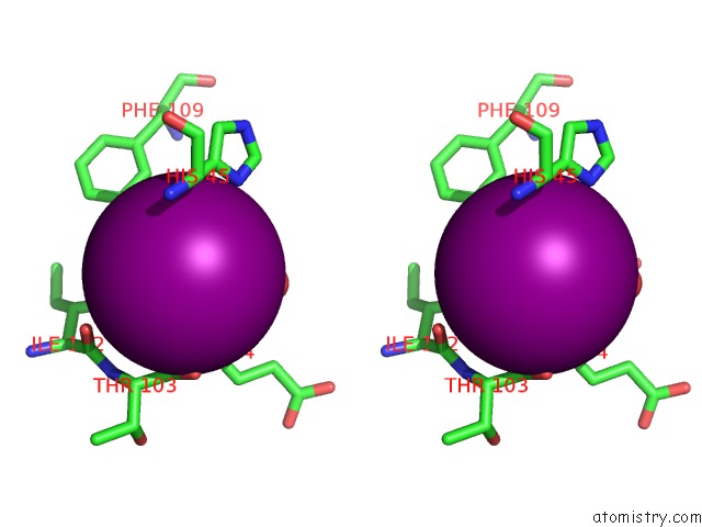
Stereo pair view
Mono view
Stereo pair view
A full contact list of Iodine with other atoms in the I binding
site number 1 of X-Ray Crystal Structure of Beta Secretase Complexed with Compound 5 within 5.0Å range:
|
Iodine binding site 2 out of 3 in 2of0
Go back to
Iodine binding site 2 out
of 3 in the X-Ray Crystal Structure of Beta Secretase Complexed with Compound 5
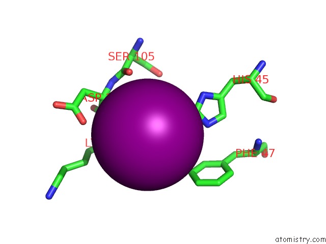
Mono view
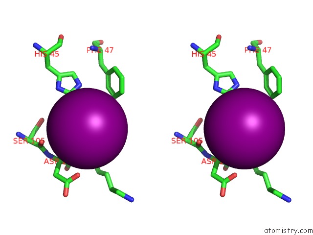
Stereo pair view
Mono view
Stereo pair view
A full contact list of Iodine with other atoms in the I binding
site number 2 of X-Ray Crystal Structure of Beta Secretase Complexed with Compound 5 within 5.0Å range:
|
Iodine binding site 3 out of 3 in 2of0
Go back to
Iodine binding site 3 out
of 3 in the X-Ray Crystal Structure of Beta Secretase Complexed with Compound 5
Mono view
Stereo pair view
Mono view
Stereo pair view
A full contact list of Iodine with other atoms in the I binding
site number 3 of X-Ray Crystal Structure of Beta Secretase Complexed with Compound 5 within 5.0Å range:
|
Reference:
C.W.Murray,
O.Callaghan,
G.Chessari,
A.Cleasby,
M.Congreve,
M.Frederickson,
M.J.Hartshorn,
R.Mcmenamin,
S.Patel,
N.Wallis.
Application of Fragment Screening By X-Ray Crystallography to Beta-Secretase. J.Med.Chem. V. 50 1116 2007.
ISSN: ISSN 0022-2623
PubMed: 17315856
DOI: 10.1021/JM0611962
Page generated: Sun Aug 11 14:10:07 2024
ISSN: ISSN 0022-2623
PubMed: 17315856
DOI: 10.1021/JM0611962
Last articles
Zn in 9J0NZn in 9J0O
Zn in 9J0P
Zn in 9FJX
Zn in 9EKB
Zn in 9C0F
Zn in 9CAH
Zn in 9CH0
Zn in 9CH3
Zn in 9CH1