Iodine »
PDB 2nqx-2qpy »
2q2l »
Iodine in PDB 2q2l: Crystal Structure of Superoxide Dismutase From P. Atrosanguina
Enzymatic activity of Crystal Structure of Superoxide Dismutase From P. Atrosanguina
All present enzymatic activity of Crystal Structure of Superoxide Dismutase From P. Atrosanguina:
1.15.1.1;
1.15.1.1;
Protein crystallography data
The structure of Crystal Structure of Superoxide Dismutase From P. Atrosanguina, PDB code: 2q2l
was solved by
Y.Manickam,
J.Gill,
P.C.Mishra,
A.Sharma,
with X-Ray Crystallography technique. A brief refinement statistics is given in the table below:
| Resolution Low / High (Å) | 50.00 / 2.37 |
| Space group | C 1 2 1 |
| Cell size a, b, c (Å), α, β, γ (°) | 89.140, 64.120, 63.620, 90.00, 124.33, 90.00 |
| R / Rfree (%) | 16.7 / 23.4 |
Other elements in 2q2l:
The structure of Crystal Structure of Superoxide Dismutase From P. Atrosanguina also contains other interesting chemical elements:
| Zinc | (Zn) | 2 atoms |
Iodine Binding Sites:
The binding sites of Iodine atom in the Crystal Structure of Superoxide Dismutase From P. Atrosanguina
(pdb code 2q2l). This binding sites where shown within
5.0 Angstroms radius around Iodine atom.
In total 10 binding sites of Iodine where determined in the Crystal Structure of Superoxide Dismutase From P. Atrosanguina, PDB code: 2q2l:
Jump to Iodine binding site number: 1; 2; 3; 4; 5; 6; 7; 8; 9; 10;
In total 10 binding sites of Iodine where determined in the Crystal Structure of Superoxide Dismutase From P. Atrosanguina, PDB code: 2q2l:
Jump to Iodine binding site number: 1; 2; 3; 4; 5; 6; 7; 8; 9; 10;
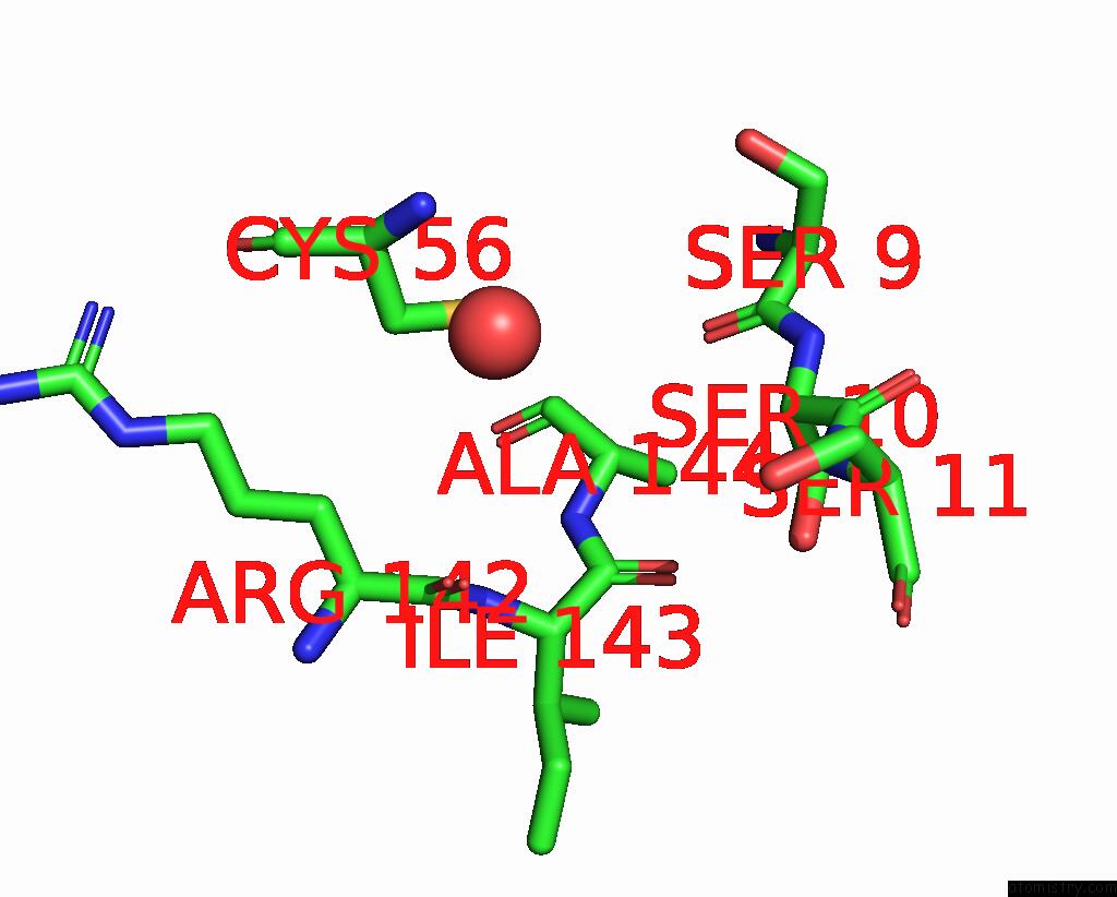
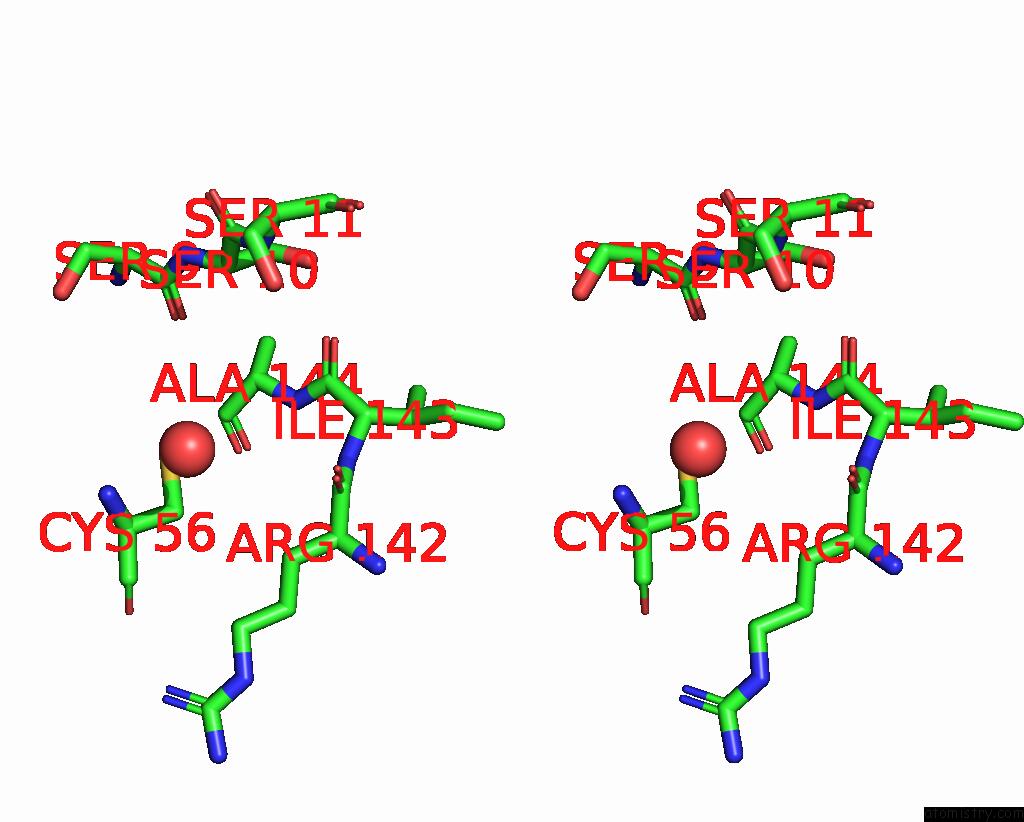
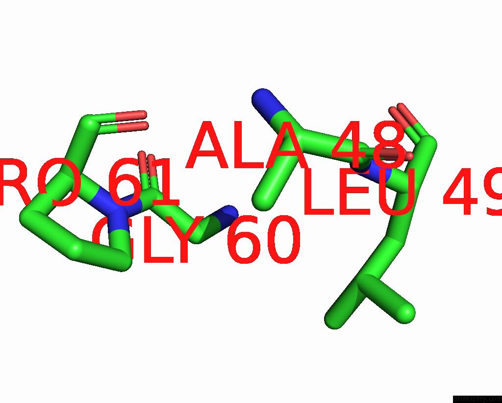
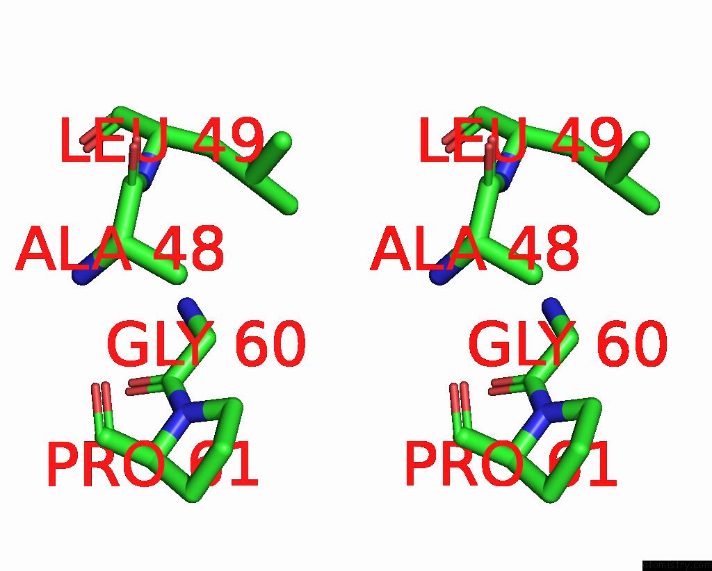
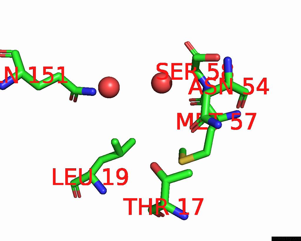
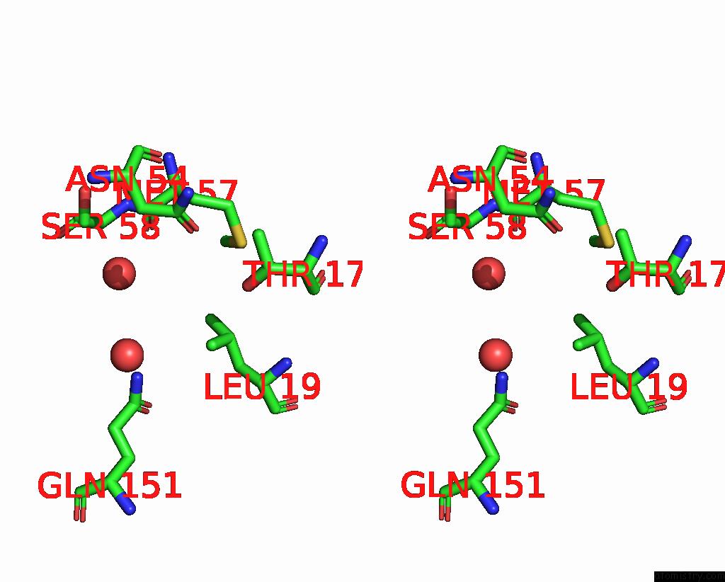
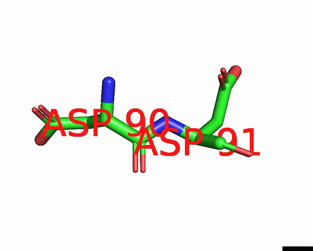
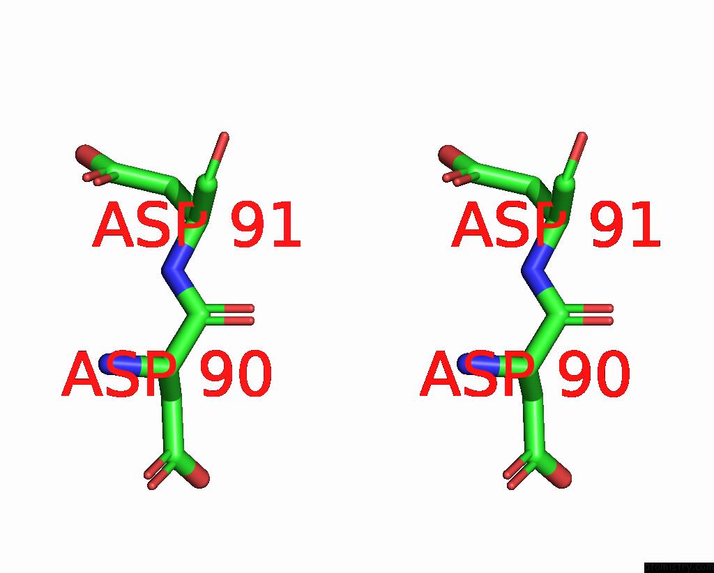
Iodine binding site 1 out of 10 in 2q2l
Go back to
Iodine binding site 1 out
of 10 in the Crystal Structure of Superoxide Dismutase From P. Atrosanguina
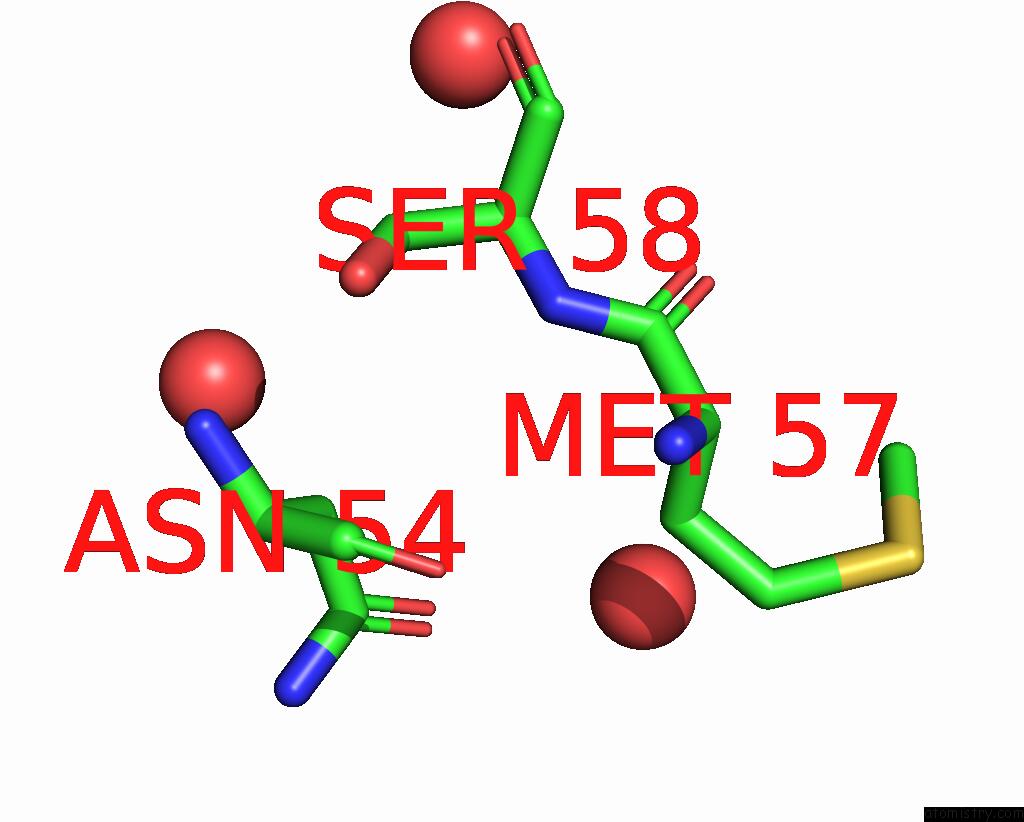
Mono view
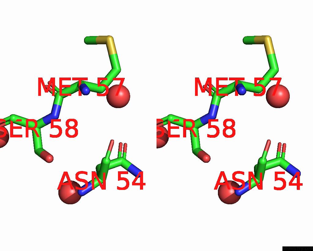
Stereo pair view
Mono view
Stereo pair view
A full contact list of Iodine with other atoms in the I binding
site number 1 of Crystal Structure of Superoxide Dismutase From P. Atrosanguina within 5.0Å range:
|
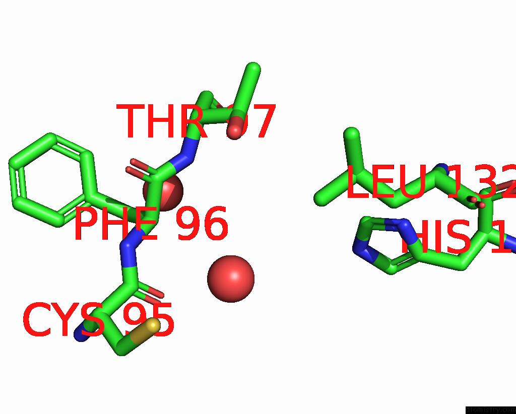
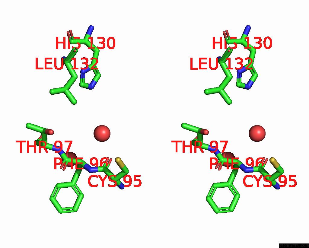
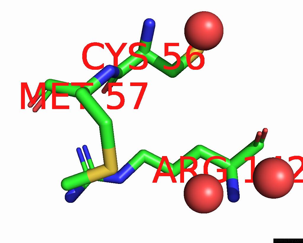
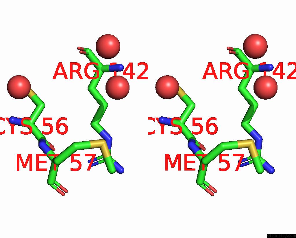
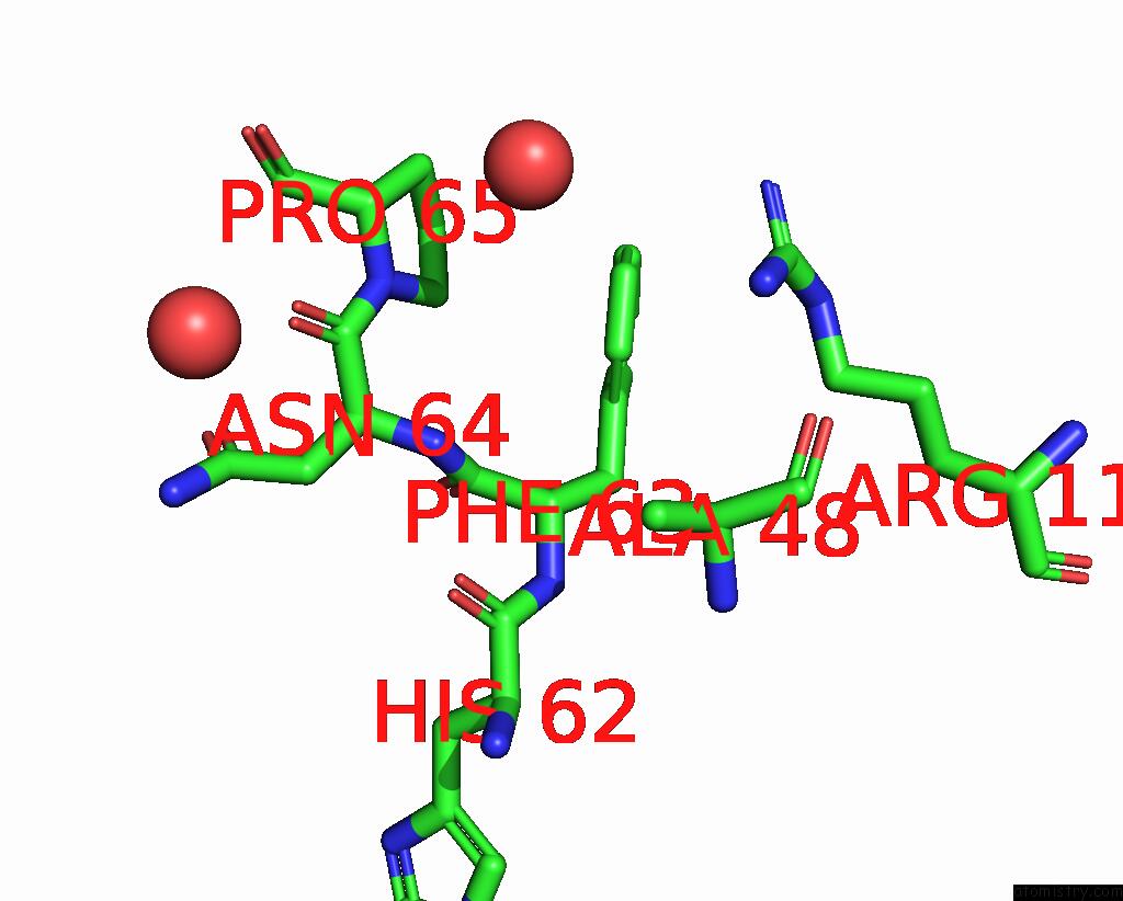
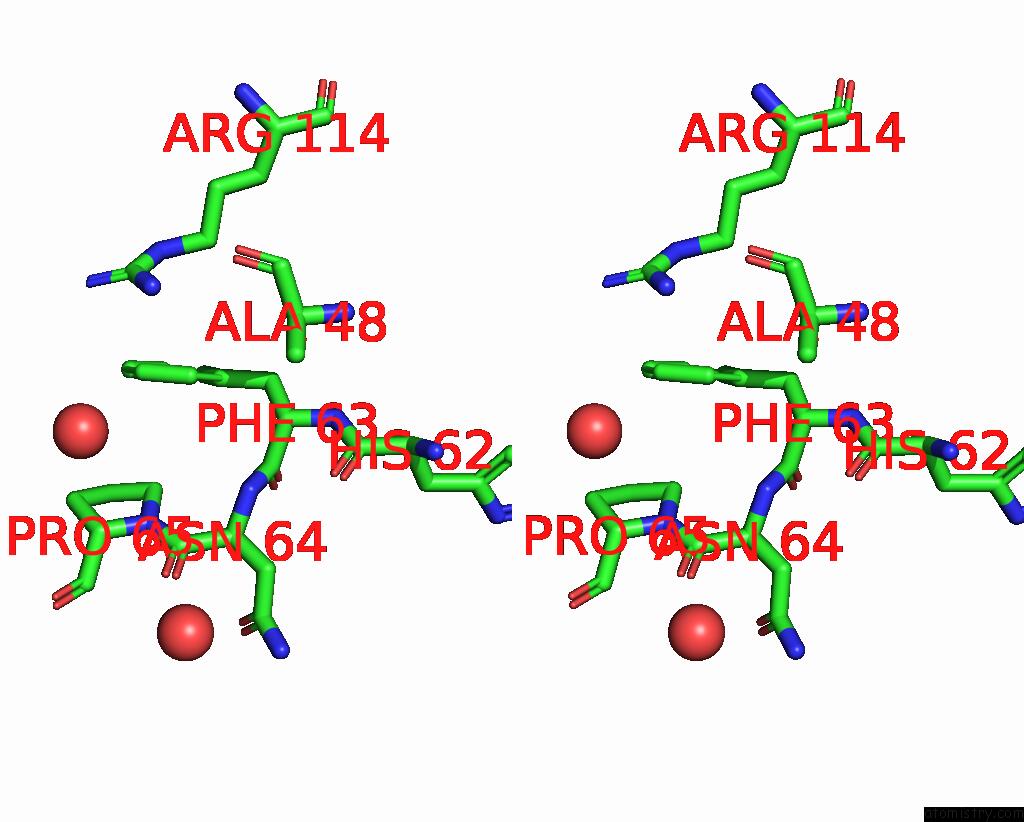
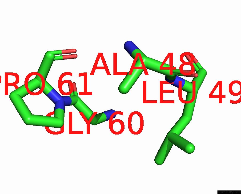
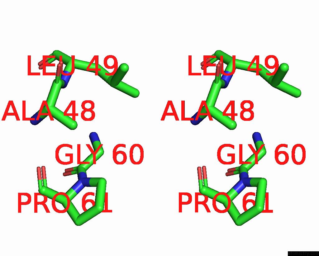
Iodine binding site 2 out of 10 in 2q2l
Go back to
Iodine binding site 2 out
of 10 in the Crystal Structure of Superoxide Dismutase From P. Atrosanguina
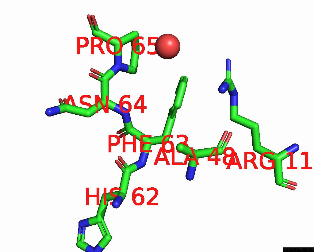
Mono view
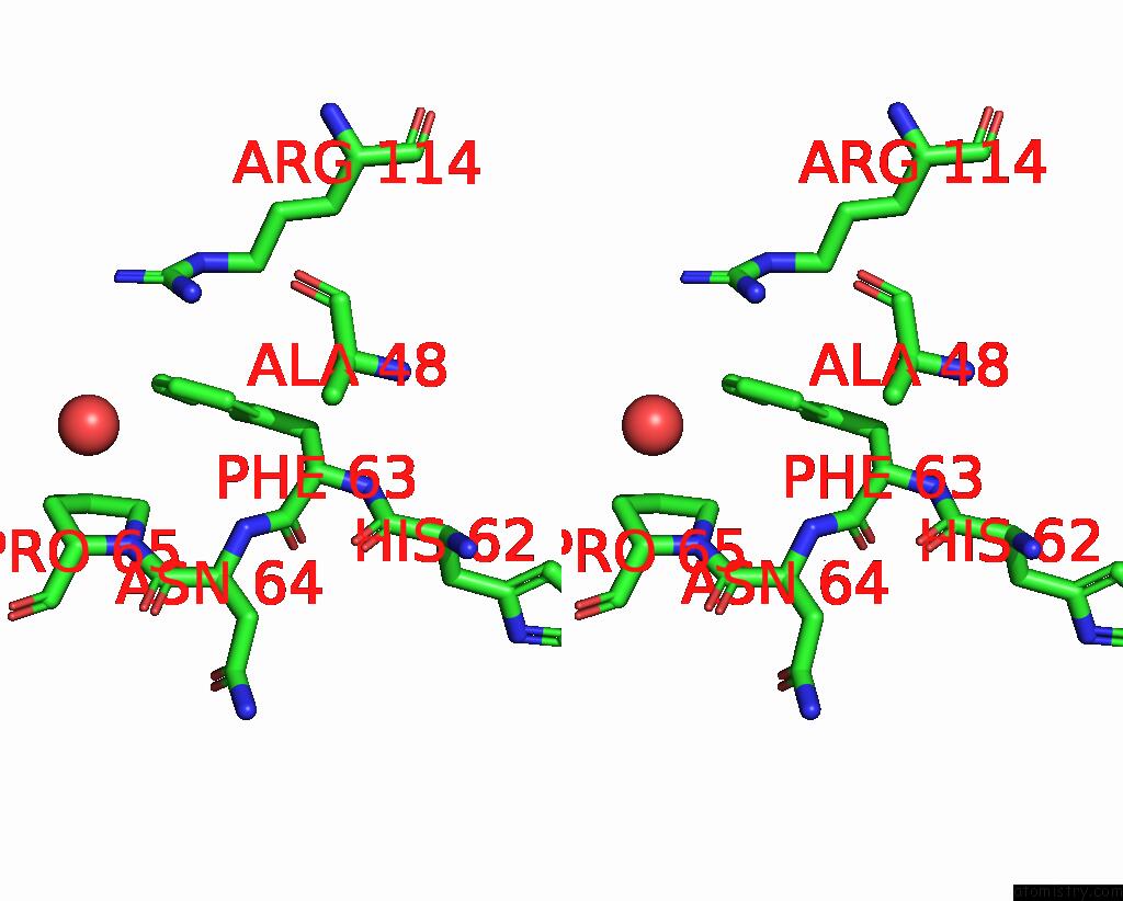
Stereo pair view
Mono view
Stereo pair view
A full contact list of Iodine with other atoms in the I binding
site number 2 of Crystal Structure of Superoxide Dismutase From P. Atrosanguina within 5.0Å range:
|
Iodine binding site 3 out of 10 in 2q2l
Go back to
Iodine binding site 3 out
of 10 in the Crystal Structure of Superoxide Dismutase From P. Atrosanguina
Mono view
Stereo pair view
Mono view
Stereo pair view
A full contact list of Iodine with other atoms in the I binding
site number 3 of Crystal Structure of Superoxide Dismutase From P. Atrosanguina within 5.0Å range:
|
Iodine binding site 4 out of 10 in 2q2l
Go back to
Iodine binding site 4 out
of 10 in the Crystal Structure of Superoxide Dismutase From P. Atrosanguina
Mono view
Stereo pair view
Mono view
Stereo pair view
A full contact list of Iodine with other atoms in the I binding
site number 4 of Crystal Structure of Superoxide Dismutase From P. Atrosanguina within 5.0Å range:
|
Iodine binding site 5 out of 10 in 2q2l
Go back to
Iodine binding site 5 out
of 10 in the Crystal Structure of Superoxide Dismutase From P. Atrosanguina
Mono view
Stereo pair view
Mono view
Stereo pair view
A full contact list of Iodine with other atoms in the I binding
site number 5 of Crystal Structure of Superoxide Dismutase From P. Atrosanguina within 5.0Å range:
|
Iodine binding site 6 out of 10 in 2q2l
Go back to
Iodine binding site 6 out
of 10 in the Crystal Structure of Superoxide Dismutase From P. Atrosanguina
Mono view
Stereo pair view
Mono view
Stereo pair view
A full contact list of Iodine with other atoms in the I binding
site number 6 of Crystal Structure of Superoxide Dismutase From P. Atrosanguina within 5.0Å range:
|
Iodine binding site 7 out of 10 in 2q2l
Go back to
Iodine binding site 7 out
of 10 in the Crystal Structure of Superoxide Dismutase From P. Atrosanguina
Mono view
Stereo pair view
Mono view
Stereo pair view
A full contact list of Iodine with other atoms in the I binding
site number 7 of Crystal Structure of Superoxide Dismutase From P. Atrosanguina within 5.0Å range:
|
Iodine binding site 8 out of 10 in 2q2l
Go back to
Iodine binding site 8 out
of 10 in the Crystal Structure of Superoxide Dismutase From P. Atrosanguina
Mono view
Stereo pair view
Mono view
Stereo pair view
A full contact list of Iodine with other atoms in the I binding
site number 8 of Crystal Structure of Superoxide Dismutase From P. Atrosanguina within 5.0Å range:
|
Iodine binding site 9 out of 10 in 2q2l
Go back to
Iodine binding site 9 out
of 10 in the Crystal Structure of Superoxide Dismutase From P. Atrosanguina
Mono view
Stereo pair view
Mono view
Stereo pair view
A full contact list of Iodine with other atoms in the I binding
site number 9 of Crystal Structure of Superoxide Dismutase From P. Atrosanguina within 5.0Å range:
|
Iodine binding site 10 out of 10 in 2q2l
Go back to
Iodine binding site 10 out
of 10 in the Crystal Structure of Superoxide Dismutase From P. Atrosanguina
Mono view
Stereo pair view
Mono view
Stereo pair view
A full contact list of Iodine with other atoms in the I binding
site number 10 of Crystal Structure of Superoxide Dismutase From P. Atrosanguina within 5.0Å range:
|
Reference:
M.Yogavel,
J.Gill,
P.C.Mishra,
A.Sharma.
Sad Phasing of A Structure Based on Cocrystallized Iodides Using An in-House Cu Kalpha X-Ray Source: Effects of Data Redundancy and Completeness on Structure Solution Acta Crystallogr.,Sect.D V. 63 931 2007.
ISSN: ISSN 0907-4449
PubMed: 17642520
DOI: 10.1107/S0907444907029174
Page generated: Sun Aug 11 14:18:18 2024
ISSN: ISSN 0907-4449
PubMed: 17642520
DOI: 10.1107/S0907444907029174
Last articles
Zn in 9MJ5Zn in 9HNW
Zn in 9G0L
Zn in 9FNE
Zn in 9DZN
Zn in 9E0I
Zn in 9D32
Zn in 9DAK
Zn in 8ZXC
Zn in 8ZUF