Iodine »
PDB 2qqt-2y5e »
2vma »
Iodine in PDB 2vma: The Three-Dimensional Structure of the Cytoplasmic Domains of Epsf From the Type 2 Secretion System of Vibrio Cholerae
Protein crystallography data
The structure of The Three-Dimensional Structure of the Cytoplasmic Domains of Epsf From the Type 2 Secretion System of Vibrio Cholerae, PDB code: 2vma
was solved by
J.Abendroth,
K.V.Korotkov,
D.D.Mitchell,
A.Kreger,
W.G.J.Hol,
with X-Ray Crystallography technique. A brief refinement statistics is given in the table below:
| Resolution Low / High (Å) | 46.32 / 1.90 |
| Space group | P 21 21 21 |
| Cell size a, b, c (Å), α, β, γ (°) | 48.816, 54.368, 88.587, 90.00, 90.00, 90.00 |
| R / Rfree (%) | 20.8 / 25.5 |
Other elements in 2vma:
The structure of The Three-Dimensional Structure of the Cytoplasmic Domains of Epsf From the Type 2 Secretion System of Vibrio Cholerae also contains other interesting chemical elements:
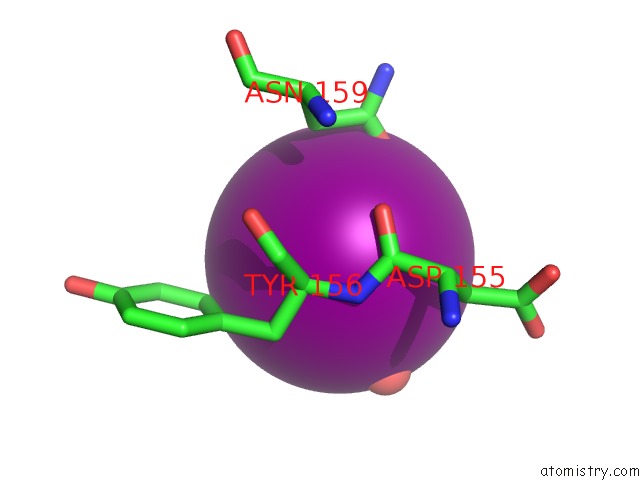
| Calcium | (Ca) | 3 atoms |
Iodine Binding Sites:
Pages:
>>> Page 1 <<< Page 2, Binding sites: 11 - 15;Binding sites:
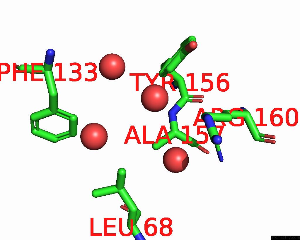
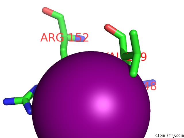
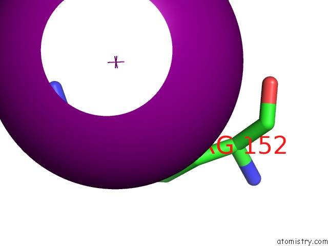
The binding sites of Iodine atom in the The Three-Dimensional Structure of the Cytoplasmic Domains of Epsf From the Type 2 Secretion System of Vibrio Cholerae (pdb code 2vma). This binding sites where shown within 5.0 Angstroms radius around Iodine atom.In total 15 binding sites of Iodine where determined in the The Three-Dimensional Structure of the Cytoplasmic Domains of Epsf From the Type 2 Secretion System of Vibrio Cholerae, PDB code: 2vma:
Jump to Iodine binding site number: 1; 2; 3; 4; 5; 6; 7; 8; 9; 10;
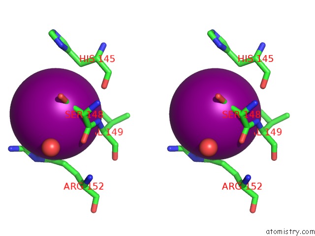
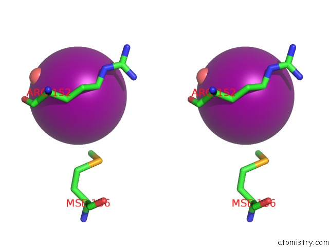
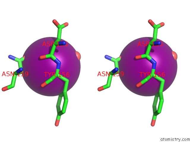
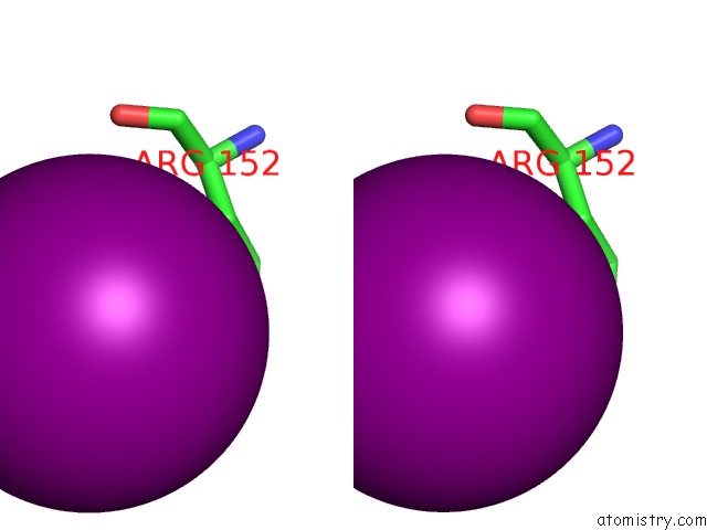
Iodine binding site 1 out of 15 in 2vma
Go back to
Iodine binding site 1 out
of 15 in the The Three-Dimensional Structure of the Cytoplasmic Domains of Epsf From the Type 2 Secretion System of Vibrio Cholerae
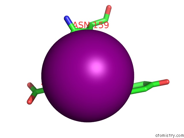
Mono view
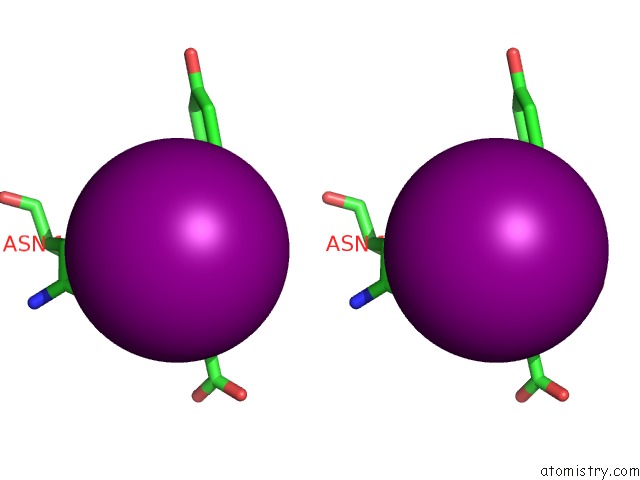
Stereo pair view
Mono view
Stereo pair view
A full contact list of Iodine with other atoms in the I binding
site number 1 of The Three-Dimensional Structure of the Cytoplasmic Domains of Epsf From the Type 2 Secretion System of Vibrio Cholerae within 5.0Å range:
|
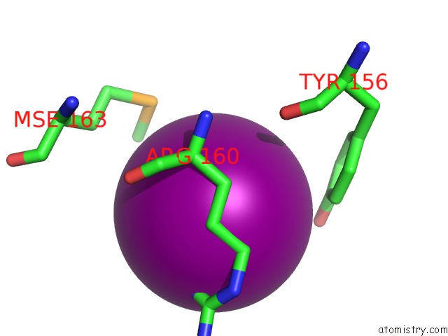
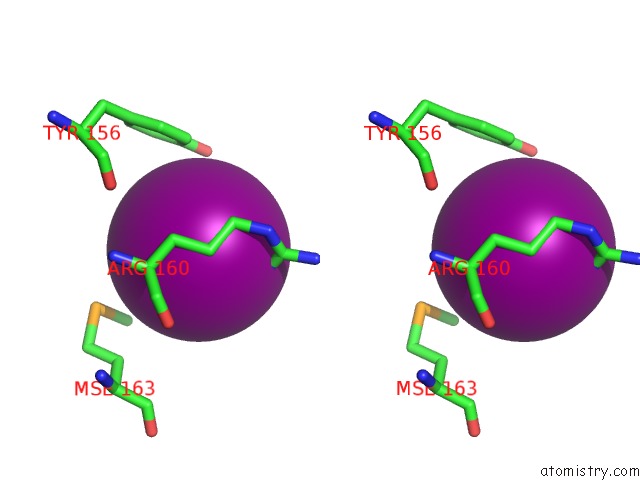
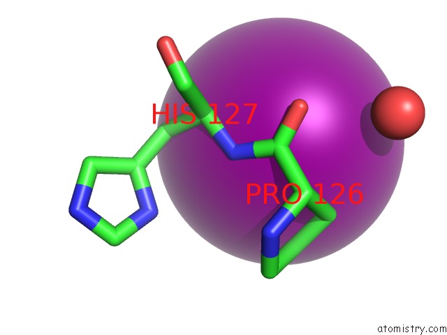
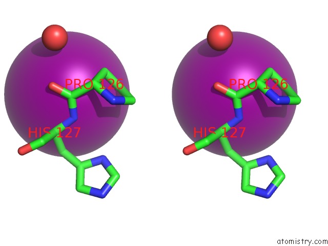
Iodine binding site 2 out of 15 in 2vma
Go back to
Iodine binding site 2 out
of 15 in the The Three-Dimensional Structure of the Cytoplasmic Domains of Epsf From the Type 2 Secretion System of Vibrio Cholerae
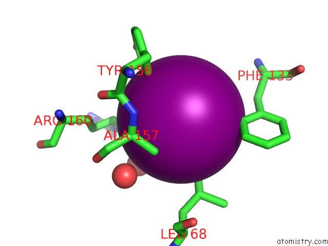
Mono view

Stereo pair view
Mono view
Stereo pair view
A full contact list of Iodine with other atoms in the I binding
site number 2 of The Three-Dimensional Structure of the Cytoplasmic Domains of Epsf From the Type 2 Secretion System of Vibrio Cholerae within 5.0Å range:
|
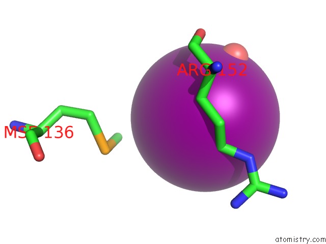
Iodine binding site 3 out of 15 in 2vma
Go back to
Iodine binding site 3 out
of 15 in the The Three-Dimensional Structure of the Cytoplasmic Domains of Epsf From the Type 2 Secretion System of Vibrio Cholerae
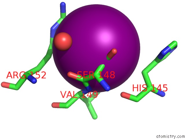
Mono view
Stereo pair view
Mono view
Stereo pair view
A full contact list of Iodine with other atoms in the I binding
site number 3 of The Three-Dimensional Structure of the Cytoplasmic Domains of Epsf From the Type 2 Secretion System of Vibrio Cholerae within 5.0Å range:
|
Iodine binding site 4 out of 15 in 2vma
Go back to
Iodine binding site 4 out
of 15 in the The Three-Dimensional Structure of the Cytoplasmic Domains of Epsf From the Type 2 Secretion System of Vibrio Cholerae
Mono view
Stereo pair view
Mono view
Stereo pair view
A full contact list of Iodine with other atoms in the I binding
site number 4 of The Three-Dimensional Structure of the Cytoplasmic Domains of Epsf From the Type 2 Secretion System of Vibrio Cholerae within 5.0Å range:
|
Iodine binding site 5 out of 15 in 2vma
Go back to
Iodine binding site 5 out
of 15 in the The Three-Dimensional Structure of the Cytoplasmic Domains of Epsf From the Type 2 Secretion System of Vibrio Cholerae
Mono view
Stereo pair view
Mono view
Stereo pair view
A full contact list of Iodine with other atoms in the I binding
site number 5 of The Three-Dimensional Structure of the Cytoplasmic Domains of Epsf From the Type 2 Secretion System of Vibrio Cholerae within 5.0Å range:
|
Iodine binding site 6 out of 15 in 2vma
Go back to
Iodine binding site 6 out
of 15 in the The Three-Dimensional Structure of the Cytoplasmic Domains of Epsf From the Type 2 Secretion System of Vibrio Cholerae
Mono view
Stereo pair view
Mono view
Stereo pair view
A full contact list of Iodine with other atoms in the I binding
site number 6 of The Three-Dimensional Structure of the Cytoplasmic Domains of Epsf From the Type 2 Secretion System of Vibrio Cholerae within 5.0Å range:
|
Iodine binding site 7 out of 15 in 2vma
Go back to
Iodine binding site 7 out
of 15 in the The Three-Dimensional Structure of the Cytoplasmic Domains of Epsf From the Type 2 Secretion System of Vibrio Cholerae
Mono view
Stereo pair view
Mono view
Stereo pair view
A full contact list of Iodine with other atoms in the I binding
site number 7 of The Three-Dimensional Structure of the Cytoplasmic Domains of Epsf From the Type 2 Secretion System of Vibrio Cholerae within 5.0Å range:
|
Iodine binding site 8 out of 15 in 2vma
Go back to
Iodine binding site 8 out
of 15 in the The Three-Dimensional Structure of the Cytoplasmic Domains of Epsf From the Type 2 Secretion System of Vibrio Cholerae
Mono view

Stereo pair view
Mono view
Stereo pair view
A full contact list of Iodine with other atoms in the I binding
site number 8 of The Three-Dimensional Structure of the Cytoplasmic Domains of Epsf From the Type 2 Secretion System of Vibrio Cholerae within 5.0Å range:
|
Iodine binding site 9 out of 15 in 2vma
Go back to
Iodine binding site 9 out
of 15 in the The Three-Dimensional Structure of the Cytoplasmic Domains of Epsf From the Type 2 Secretion System of Vibrio Cholerae
Mono view

Stereo pair view
Mono view
Stereo pair view
A full contact list of Iodine with other atoms in the I binding
site number 9 of The Three-Dimensional Structure of the Cytoplasmic Domains of Epsf From the Type 2 Secretion System of Vibrio Cholerae within 5.0Å range:
|
Iodine binding site 10 out of 15 in 2vma
Go back to
Iodine binding site 10 out
of 15 in the The Three-Dimensional Structure of the Cytoplasmic Domains of Epsf From the Type 2 Secretion System of Vibrio Cholerae
Mono view
Stereo pair view
Mono view
Stereo pair view
A full contact list of Iodine with other atoms in the I binding
site number 10 of The Three-Dimensional Structure of the Cytoplasmic Domains of Epsf From the Type 2 Secretion System of Vibrio Cholerae within 5.0Å range:
|
Reference:
J.Abendroth,
D.D.Mitchell,
K.V.Korotkov,
T.L.Johnson,
A.Kreger,
M.Sandkvist,
W.G.Hol.
The Three-Dimensional Structure of the Cytoplasmic Domains of Epsf From the Type 2 Secretion System of Vibrio Cholerae. J.Struct.Biol. V. 166 303 2009.
ISSN: ISSN 1047-8477
PubMed: 19324092
DOI: 10.1016/J.JSB.2009.03.009
Page generated: Fri Aug 8 13:38:19 2025
ISSN: ISSN 1047-8477
PubMed: 19324092
DOI: 10.1016/J.JSB.2009.03.009
Last articles
I in 4AGQI in 4AGP
I in 4AGO
I in 4AGM
I in 4AGL
I in 4AGN
I in 464D
I in 4A3P
I in 444D
I in 445D