Iodine »
PDB 2qqt-2y5e »
2w86 »
Iodine in PDB 2w86: Crystal Structure of Fibrillin-1 Domains CBEGF9HYB2CBEGF10, Calcium Saturated Form
Protein crystallography data
The structure of Crystal Structure of Fibrillin-1 Domains CBEGF9HYB2CBEGF10, Calcium Saturated Form, PDB code: 2w86
was solved by
S.A.Jensen,
S.Iqbal,
E.D.Lowe,
C.Redfield,
P.A.Handford,
with X-Ray Crystallography technique. A brief refinement statistics is given in the table below:
| Resolution Low / High (Å) | 41.750 / 1.80 |
| Space group | P 21 21 21 |
| Cell size a, b, c (Å), α, β, γ (°) | 31.232, 47.258, 89.105, 90.00, 90.00, 90.00 |
| R / Rfree (%) | 18.51 / 22.12 |
Other elements in 2w86:
The structure of Crystal Structure of Fibrillin-1 Domains CBEGF9HYB2CBEGF10, Calcium Saturated Form also contains other interesting chemical elements:
| Calcium | (Ca) | 2 atoms |
Iodine Binding Sites:
The binding sites of Iodine atom in the Crystal Structure of Fibrillin-1 Domains CBEGF9HYB2CBEGF10, Calcium Saturated Form
(pdb code 2w86). This binding sites where shown within
5.0 Angstroms radius around Iodine atom.
In total 3 binding sites of Iodine where determined in the Crystal Structure of Fibrillin-1 Domains CBEGF9HYB2CBEGF10, Calcium Saturated Form, PDB code: 2w86:
Jump to Iodine binding site number: 1; 2; 3;
In total 3 binding sites of Iodine where determined in the Crystal Structure of Fibrillin-1 Domains CBEGF9HYB2CBEGF10, Calcium Saturated Form, PDB code: 2w86:
Jump to Iodine binding site number: 1; 2; 3;
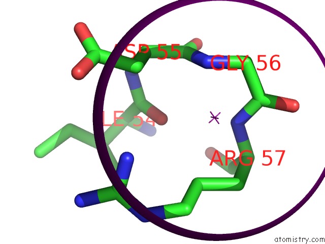
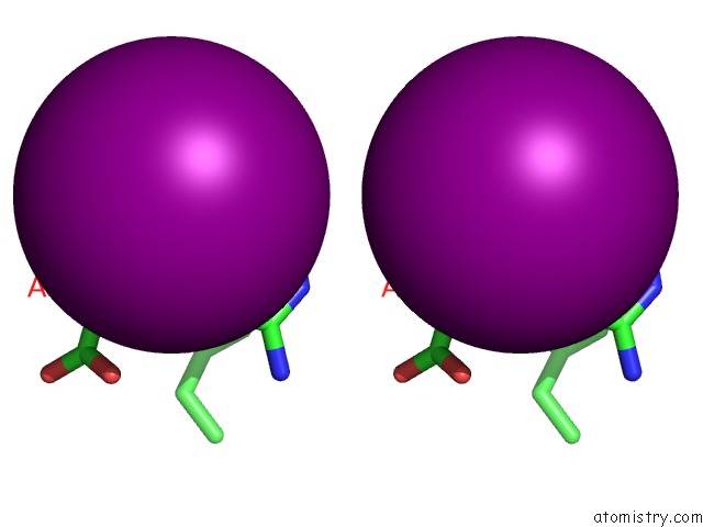
Iodine binding site 1 out of 3 in 2w86
Go back to
Iodine binding site 1 out
of 3 in the Crystal Structure of Fibrillin-1 Domains CBEGF9HYB2CBEGF10, Calcium Saturated Form
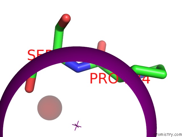
Mono view
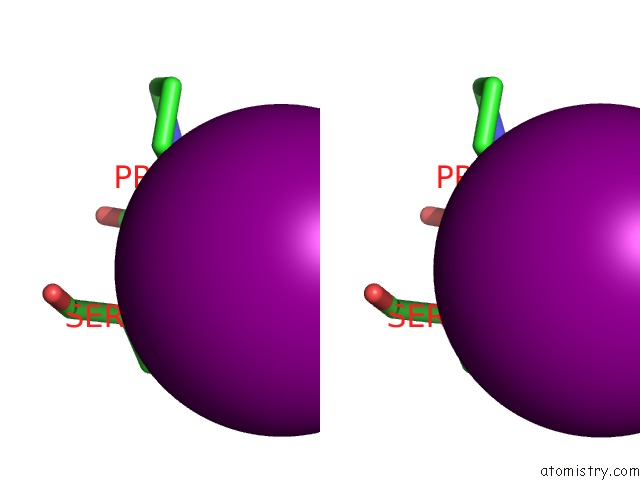
Stereo pair view
Mono view
Stereo pair view
A full contact list of Iodine with other atoms in the I binding
site number 1 of Crystal Structure of Fibrillin-1 Domains CBEGF9HYB2CBEGF10, Calcium Saturated Form within 5.0Å range:
|
Iodine binding site 2 out of 3 in 2w86
Go back to
Iodine binding site 2 out
of 3 in the Crystal Structure of Fibrillin-1 Domains CBEGF9HYB2CBEGF10, Calcium Saturated Form
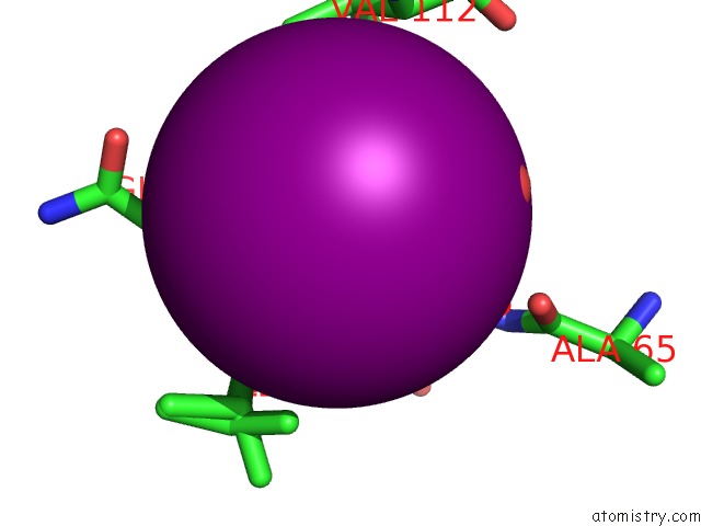
Mono view
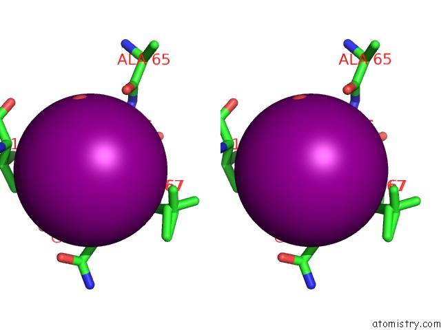
Stereo pair view
Mono view
Stereo pair view
A full contact list of Iodine with other atoms in the I binding
site number 2 of Crystal Structure of Fibrillin-1 Domains CBEGF9HYB2CBEGF10, Calcium Saturated Form within 5.0Å range:
|
Iodine binding site 3 out of 3 in 2w86
Go back to
Iodine binding site 3 out
of 3 in the Crystal Structure of Fibrillin-1 Domains CBEGF9HYB2CBEGF10, Calcium Saturated Form
Mono view
Stereo pair view
Mono view
Stereo pair view
A full contact list of Iodine with other atoms in the I binding
site number 3 of Crystal Structure of Fibrillin-1 Domains CBEGF9HYB2CBEGF10, Calcium Saturated Form within 5.0Å range:
|
Reference:
S.A.Jensen,
S.Iqbal,
E.D.Lowe,
C.Redfield,
P.A.Handford.
Structure and Interdomain Interactions of A Hybrid Domain: A Disulphide-Rich Module of the Fibrillin/Ltbp Superfamily of Matrix Proteins. Structure V. 17 759 2009.
ISSN: ISSN 0969-2126
PubMed: 19446531
DOI: 10.1016/J.STR.2009.03.014
Page generated: Fri Aug 8 13:39:28 2025
ISSN: ISSN 0969-2126
PubMed: 19446531
DOI: 10.1016/J.STR.2009.03.014
Last articles
K in 4EVYK in 4EOU
K in 4ETM
K in 4ESK
K in 4ES8
K in 4ERT
K in 4ERD
K in 4ENC
K in 4EK1
K in 4ENB