Iodine »
PDB 2qqt-2y5e »
2wko »
Iodine in PDB 2wko: Structure of Metal Loaded Pathogenic SOD1 Mutant G93A.
Enzymatic activity of Structure of Metal Loaded Pathogenic SOD1 Mutant G93A.
All present enzymatic activity of Structure of Metal Loaded Pathogenic SOD1 Mutant G93A.:
1.15.1.1;
1.15.1.1;
Protein crystallography data
The structure of Structure of Metal Loaded Pathogenic SOD1 Mutant G93A., PDB code: 2wko
was solved by
S.V.Antonyuk,
A.Galaleldeen,
R.Strange,
L.Whitson,
N.Narayana,
A.Taylor,
J.P.Schuermann,
S.P.Holloway,
S.S.Hasnain,
P.J.Hart,
with X-Ray Crystallography technique. A brief refinement statistics is given in the table below:
| Resolution Low / High (Å) | 56.80 / 1.97 |
| Space group | P 1 21 1 |
| Cell size a, b, c (Å), α, β, γ (°) | 51.488, 47.157, 56.356, 90.00, 90.38, 90.00 |
| R / Rfree (%) | 17.36 / 23.072 |
Other elements in 2wko:
The structure of Structure of Metal Loaded Pathogenic SOD1 Mutant G93A. also contains other interesting chemical elements:
| Copper | (Cu) | 2 atoms |
| Zinc | (Zn) | 2 atoms |
Iodine Binding Sites:
The binding sites of Iodine atom in the Structure of Metal Loaded Pathogenic SOD1 Mutant G93A.
(pdb code 2wko). This binding sites where shown within
5.0 Angstroms radius around Iodine atom.
In total 4 binding sites of Iodine where determined in the Structure of Metal Loaded Pathogenic SOD1 Mutant G93A., PDB code: 2wko:
Jump to Iodine binding site number: 1; 2; 3; 4;
In total 4 binding sites of Iodine where determined in the Structure of Metal Loaded Pathogenic SOD1 Mutant G93A., PDB code: 2wko:
Jump to Iodine binding site number: 1; 2; 3; 4;
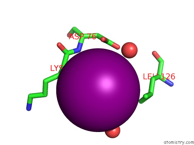
Iodine binding site 1 out of 4 in 2wko
Go back to
Iodine binding site 1 out
of 4 in the Structure of Metal Loaded Pathogenic SOD1 Mutant G93A.
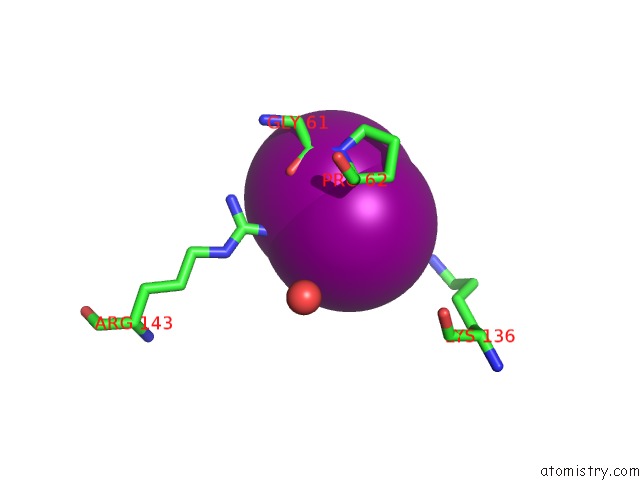
Mono view
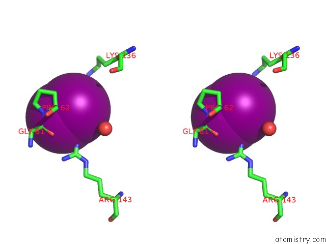
Stereo pair view
Mono view
Stereo pair view
A full contact list of Iodine with other atoms in the I binding
site number 1 of Structure of Metal Loaded Pathogenic SOD1 Mutant G93A. within 5.0Å range:
|
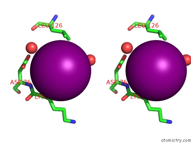
Iodine binding site 2 out of 4 in 2wko
Go back to
Iodine binding site 2 out
of 4 in the Structure of Metal Loaded Pathogenic SOD1 Mutant G93A.
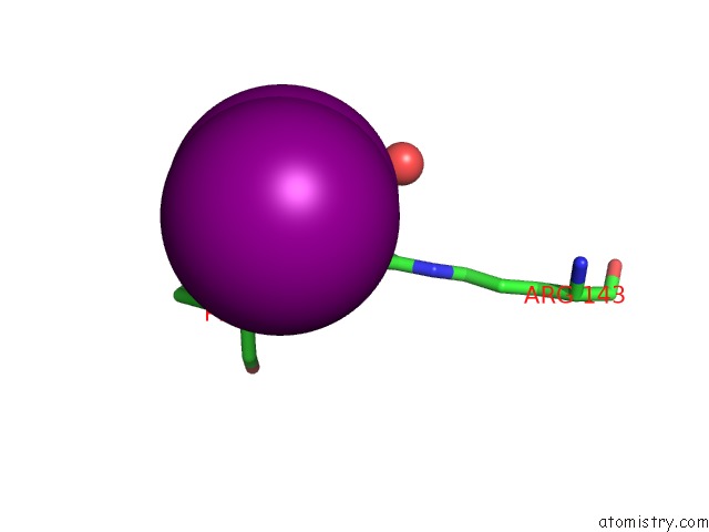
Mono view
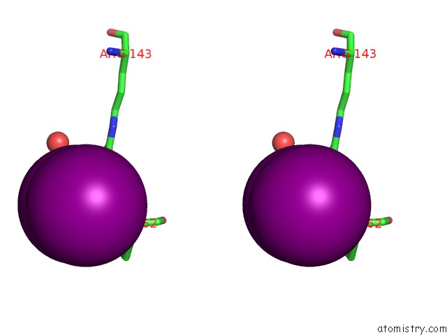
Stereo pair view
Mono view
Stereo pair view
A full contact list of Iodine with other atoms in the I binding
site number 2 of Structure of Metal Loaded Pathogenic SOD1 Mutant G93A. within 5.0Å range:
|
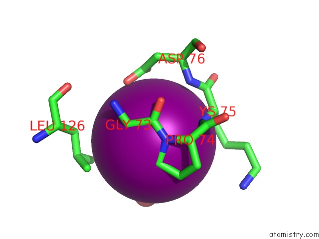
Iodine binding site 3 out of 4 in 2wko
Go back to
Iodine binding site 3 out
of 4 in the Structure of Metal Loaded Pathogenic SOD1 Mutant G93A.
Mono view
Stereo pair view
Mono view
Stereo pair view
A full contact list of Iodine with other atoms in the I binding
site number 3 of Structure of Metal Loaded Pathogenic SOD1 Mutant G93A. within 5.0Å range:
|
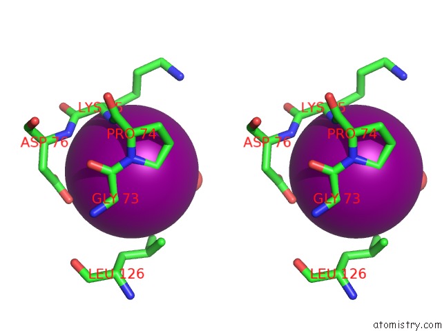
Iodine binding site 4 out of 4 in 2wko
Go back to
Iodine binding site 4 out
of 4 in the Structure of Metal Loaded Pathogenic SOD1 Mutant G93A.
Mono view
Stereo pair view
Mono view
Stereo pair view
A full contact list of Iodine with other atoms in the I binding
site number 4 of Structure of Metal Loaded Pathogenic SOD1 Mutant G93A. within 5.0Å range:
|
Reference:
A.Galaleldeen,
R.W.Strange,
L.J.Whitson,
S.V.Antonyuk,
N.Narayana,
A.B.Taylor,
J.P.Schuermann,
S.P.Holloway,
S.S.Hasnain,
P.J.Hart.
Structural and Biophysical Properties of Metal- Free Pathogenic SOD1 Mutants A4V and G93A. Arch.Biochem.Biophys. V. 492 40 2009.
ISSN: ISSN 0003-9861
PubMed: 19800308
DOI: 10.1016/J.ABB.2009.09.020
Page generated: Sun Aug 11 14:27:49 2024
ISSN: ISSN 0003-9861
PubMed: 19800308
DOI: 10.1016/J.ABB.2009.09.020
Last articles
Cl in 3UZZCl in 3UZY
Cl in 3UZW
Cl in 3UZ5
Cl in 3UXD
Cl in 3UY9
Cl in 3UZC
Cl in 3UXH
Cl in 3UXE
Cl in 3UX0