Iodine »
PDB 2qqt-2y5e »
2x76 »
Iodine in PDB 2x76: The Crystal Structure of PHAZ7 at Atomic (1.2 Angstrom) Resolution Reveals Details of the Active Site and Suggests A Substrate Binding Mode
Protein crystallography data
The structure of The Crystal Structure of PHAZ7 at Atomic (1.2 Angstrom) Resolution Reveals Details of the Active Site and Suggests A Substrate Binding Mode, PDB code: 2x76
was solved by
S.Wakadkar,
S.Hermawan,
D.Jendrossek,
A.C.Papageorgiou,
with X-Ray Crystallography technique. A brief refinement statistics is given in the table below:
| Resolution Low / High (Å) | 20.00 / 1.45 |
| Space group | P 21 21 2 |
| Cell size a, b, c (Å), α, β, γ (°) | 49.630, 140.580, 56.750, 90.00, 90.00, 90.00 |
| R / Rfree (%) | n/a / 17.8 |
Other elements in 2x76:
The structure of The Crystal Structure of PHAZ7 at Atomic (1.2 Angstrom) Resolution Reveals Details of the Active Site and Suggests A Substrate Binding Mode also contains other interesting chemical elements:
| Chlorine | (Cl) | 6 atoms |
| Sodium | (Na) | 2 atoms |
Iodine Binding Sites:
The binding sites of Iodine atom in the The Crystal Structure of PHAZ7 at Atomic (1.2 Angstrom) Resolution Reveals Details of the Active Site and Suggests A Substrate Binding Mode
(pdb code 2x76). This binding sites where shown within
5.0 Angstroms radius around Iodine atom.
In total 8 binding sites of Iodine where determined in the The Crystal Structure of PHAZ7 at Atomic (1.2 Angstrom) Resolution Reveals Details of the Active Site and Suggests A Substrate Binding Mode, PDB code: 2x76:
Jump to Iodine binding site number: 1; 2; 3; 4; 5; 6; 7; 8;
In total 8 binding sites of Iodine where determined in the The Crystal Structure of PHAZ7 at Atomic (1.2 Angstrom) Resolution Reveals Details of the Active Site and Suggests A Substrate Binding Mode, PDB code: 2x76:
Jump to Iodine binding site number: 1; 2; 3; 4; 5; 6; 7; 8;
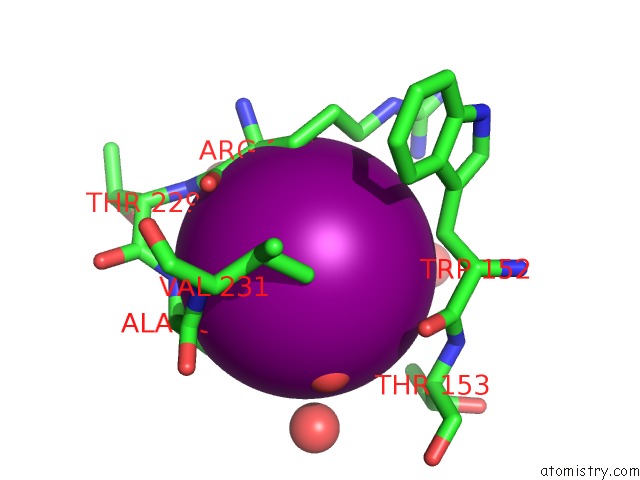
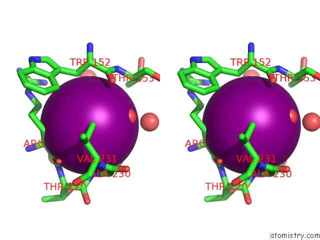
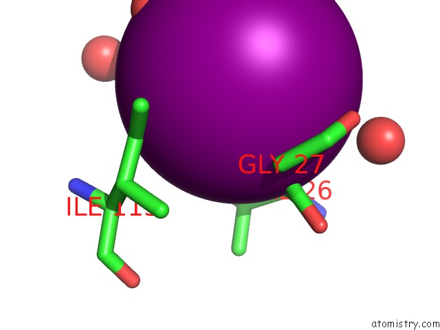
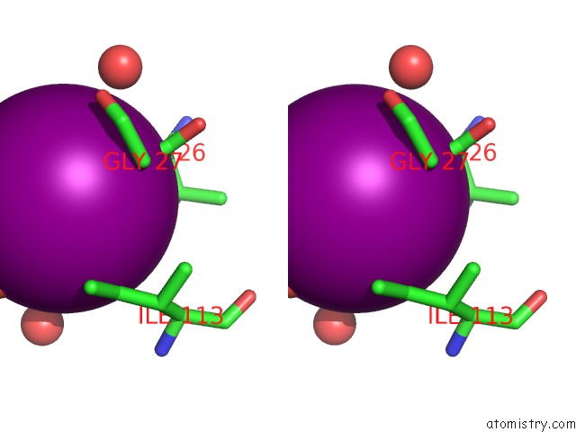
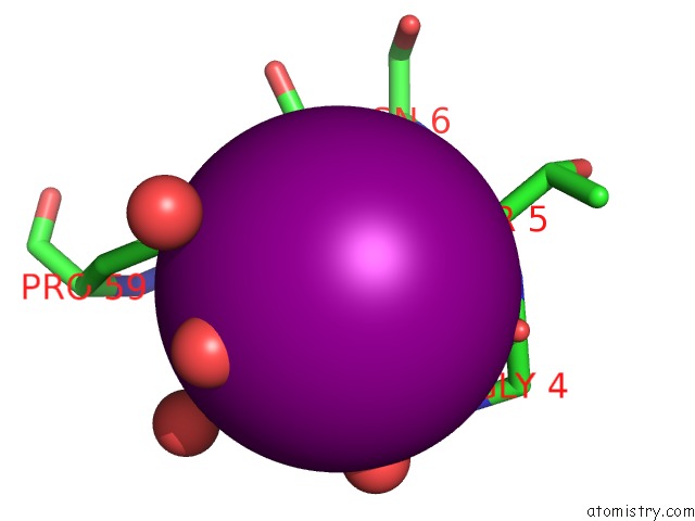
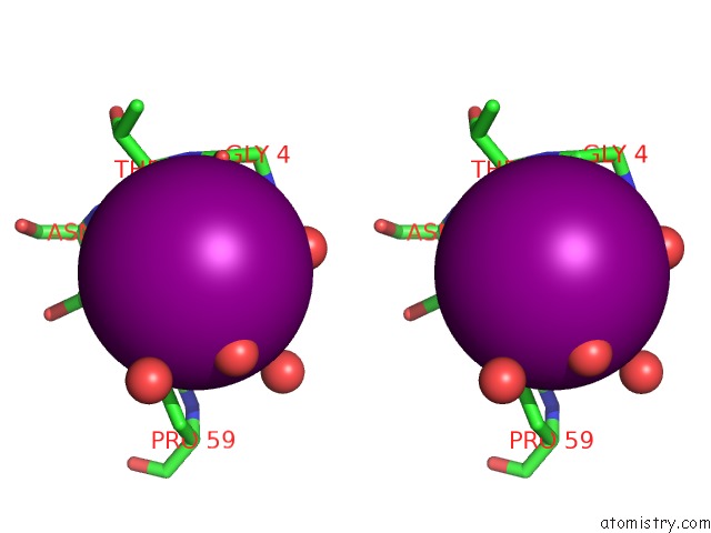
Iodine binding site 1 out of 8 in 2x76
Go back to
Iodine binding site 1 out
of 8 in the The Crystal Structure of PHAZ7 at Atomic (1.2 Angstrom) Resolution Reveals Details of the Active Site and Suggests A Substrate Binding Mode
Mono view
Stereo pair view
Mono view
Stereo pair view
A full contact list of Iodine with other atoms in the I binding
site number 1 of The Crystal Structure of PHAZ7 at Atomic (1.2 Angstrom) Resolution Reveals Details of the Active Site and Suggests A Substrate Binding Mode within 5.0Å range:
|
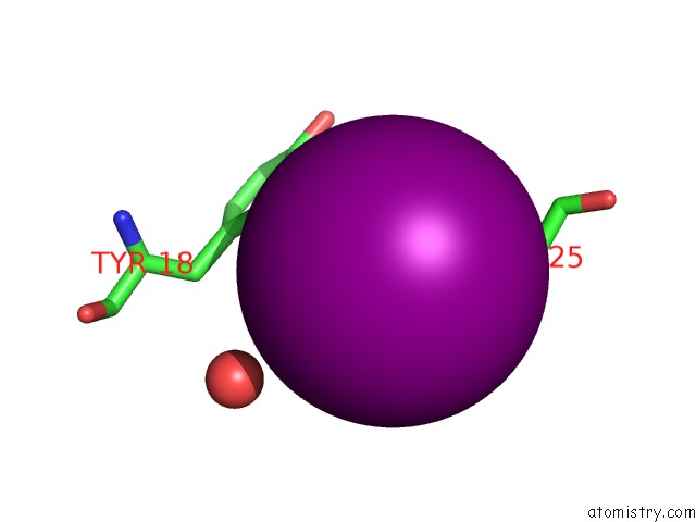
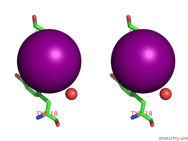
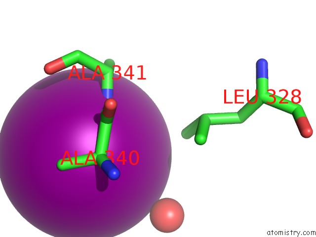
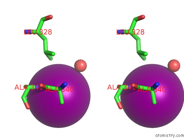
Iodine binding site 2 out of 8 in 2x76
Go back to
Iodine binding site 2 out
of 8 in the The Crystal Structure of PHAZ7 at Atomic (1.2 Angstrom) Resolution Reveals Details of the Active Site and Suggests A Substrate Binding Mode
Mono view
Stereo pair view
Mono view
Stereo pair view
A full contact list of Iodine with other atoms in the I binding
site number 2 of The Crystal Structure of PHAZ7 at Atomic (1.2 Angstrom) Resolution Reveals Details of the Active Site and Suggests A Substrate Binding Mode within 5.0Å range:
|
Iodine binding site 3 out of 8 in 2x76
Go back to
Iodine binding site 3 out
of 8 in the The Crystal Structure of PHAZ7 at Atomic (1.2 Angstrom) Resolution Reveals Details of the Active Site and Suggests A Substrate Binding Mode
Mono view
Stereo pair view
Mono view
Stereo pair view
A full contact list of Iodine with other atoms in the I binding
site number 3 of The Crystal Structure of PHAZ7 at Atomic (1.2 Angstrom) Resolution Reveals Details of the Active Site and Suggests A Substrate Binding Mode within 5.0Å range:
|
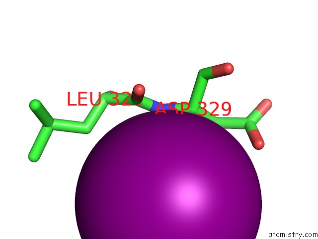
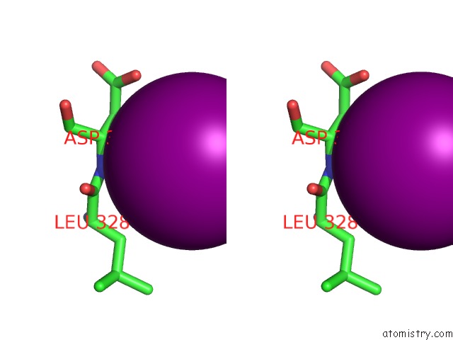
Iodine binding site 4 out of 8 in 2x76
Go back to
Iodine binding site 4 out
of 8 in the The Crystal Structure of PHAZ7 at Atomic (1.2 Angstrom) Resolution Reveals Details of the Active Site and Suggests A Substrate Binding Mode
Mono view
Stereo pair view
Mono view
Stereo pair view
A full contact list of Iodine with other atoms in the I binding
site number 4 of The Crystal Structure of PHAZ7 at Atomic (1.2 Angstrom) Resolution Reveals Details of the Active Site and Suggests A Substrate Binding Mode within 5.0Å range:
|
Iodine binding site 5 out of 8 in 2x76
Go back to
Iodine binding site 5 out
of 8 in the The Crystal Structure of PHAZ7 at Atomic (1.2 Angstrom) Resolution Reveals Details of the Active Site and Suggests A Substrate Binding Mode
Mono view
Stereo pair view
Mono view
Stereo pair view
A full contact list of Iodine with other atoms in the I binding
site number 5 of The Crystal Structure of PHAZ7 at Atomic (1.2 Angstrom) Resolution Reveals Details of the Active Site and Suggests A Substrate Binding Mode within 5.0Å range:
|
Iodine binding site 6 out of 8 in 2x76
Go back to
Iodine binding site 6 out
of 8 in the The Crystal Structure of PHAZ7 at Atomic (1.2 Angstrom) Resolution Reveals Details of the Active Site and Suggests A Substrate Binding Mode
Mono view
Stereo pair view
Mono view
Stereo pair view
A full contact list of Iodine with other atoms in the I binding
site number 6 of The Crystal Structure of PHAZ7 at Atomic (1.2 Angstrom) Resolution Reveals Details of the Active Site and Suggests A Substrate Binding Mode within 5.0Å range:
|
Iodine binding site 7 out of 8 in 2x76
Go back to
Iodine binding site 7 out
of 8 in the The Crystal Structure of PHAZ7 at Atomic (1.2 Angstrom) Resolution Reveals Details of the Active Site and Suggests A Substrate Binding Mode
Mono view
Stereo pair view
Mono view
Stereo pair view
A full contact list of Iodine with other atoms in the I binding
site number 7 of The Crystal Structure of PHAZ7 at Atomic (1.2 Angstrom) Resolution Reveals Details of the Active Site and Suggests A Substrate Binding Mode within 5.0Å range:
|
Iodine binding site 8 out of 8 in 2x76
Go back to
Iodine binding site 8 out
of 8 in the The Crystal Structure of PHAZ7 at Atomic (1.2 Angstrom) Resolution Reveals Details of the Active Site and Suggests A Substrate Binding Mode
Mono view
Stereo pair view
Mono view
Stereo pair view
A full contact list of Iodine with other atoms in the I binding
site number 8 of The Crystal Structure of PHAZ7 at Atomic (1.2 Angstrom) Resolution Reveals Details of the Active Site and Suggests A Substrate Binding Mode within 5.0Å range:
|
Reference:
S.Wakadkar,
S.Hermawan,
D.Jendrossek,
A.C.Papageorgiou.
The Structure of PHAZ7 at Atomic (1.2 A) Resolution Reveals Details of the Active Site and Suggests A Substrate-Binding Mode. Acta Crystallogr. Sect. F V. 66 648 2010STRUCT. Biol. Cryst. Commun..
ISSN: ESSN 1744-3091
PubMed: 20516591
DOI: 10.1107/S174430911001434X
Page generated: Sun Aug 11 14:28:59 2024
ISSN: ESSN 1744-3091
PubMed: 20516591
DOI: 10.1107/S174430911001434X
Last articles
Cl in 3W54Cl in 3W69
Cl in 3W33
Cl in 3W2S
Cl in 3W32
Cl in 3W2R
Cl in 3W2Q
Cl in 3W23
Cl in 3W1L
Cl in 3W2O