Iodine »
PDB 2y6q-3cgp »
2yd9 »
Iodine in PDB 2yd9: Crystal Structure of the N-Terminal IG1-3 Module of Human Receptor Protein Tyrosine Phosphatase Sigma
Enzymatic activity of Crystal Structure of the N-Terminal IG1-3 Module of Human Receptor Protein Tyrosine Phosphatase Sigma
All present enzymatic activity of Crystal Structure of the N-Terminal IG1-3 Module of Human Receptor Protein Tyrosine Phosphatase Sigma:
3.1.3.48;
3.1.3.48;
Protein crystallography data
The structure of Crystal Structure of the N-Terminal IG1-3 Module of Human Receptor Protein Tyrosine Phosphatase Sigma, PDB code: 2yd9
was solved by
C.H.Coles,
Y.Shen,
A.P.Tenney,
C.Siebold,
G.C.Sutton,
W.Lu,
J.T.Gallagher,
E.Y.Jones,
J.G.Flanagan,
A.R.Aricescu,
with X-Ray Crystallography technique. A brief refinement statistics is given in the table below:
| Resolution Low / High (Å) | 44.16 / 2.60 |
| Space group | I 2 2 2 |
| Cell size a, b, c (Å), α, β, γ (°) | 71.699, 90.043, 143.283, 90.00, 90.00, 90.00 |
| R / Rfree (%) | 24.7 / 28.9 |
Other elements in 2yd9:
The structure of Crystal Structure of the N-Terminal IG1-3 Module of Human Receptor Protein Tyrosine Phosphatase Sigma also contains other interesting chemical elements:
| Chlorine | (Cl) | 4 atoms |
Iodine Binding Sites:
The binding sites of Iodine atom in the Crystal Structure of the N-Terminal IG1-3 Module of Human Receptor Protein Tyrosine Phosphatase Sigma
(pdb code 2yd9). This binding sites where shown within
5.0 Angstroms radius around Iodine atom.
In total 4 binding sites of Iodine where determined in the Crystal Structure of the N-Terminal IG1-3 Module of Human Receptor Protein Tyrosine Phosphatase Sigma, PDB code: 2yd9:
Jump to Iodine binding site number: 1; 2; 3; 4;
In total 4 binding sites of Iodine where determined in the Crystal Structure of the N-Terminal IG1-3 Module of Human Receptor Protein Tyrosine Phosphatase Sigma, PDB code: 2yd9:
Jump to Iodine binding site number: 1; 2; 3; 4;
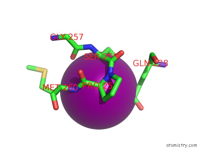
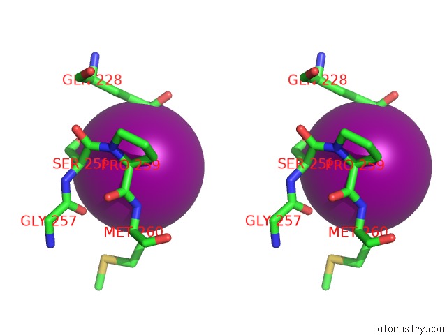
Iodine binding site 1 out of 4 in 2yd9
Go back to
Iodine binding site 1 out
of 4 in the Crystal Structure of the N-Terminal IG1-3 Module of Human Receptor Protein Tyrosine Phosphatase Sigma
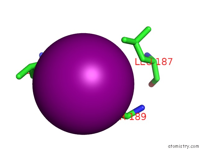
Mono view
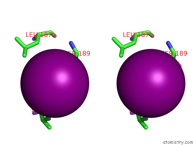
Stereo pair view
Mono view
Stereo pair view
A full contact list of Iodine with other atoms in the I binding
site number 1 of Crystal Structure of the N-Terminal IG1-3 Module of Human Receptor Protein Tyrosine Phosphatase Sigma within 5.0Å range:
|
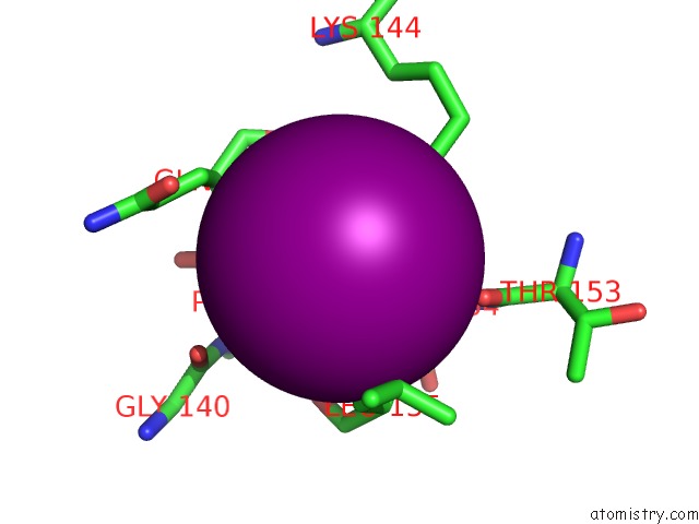
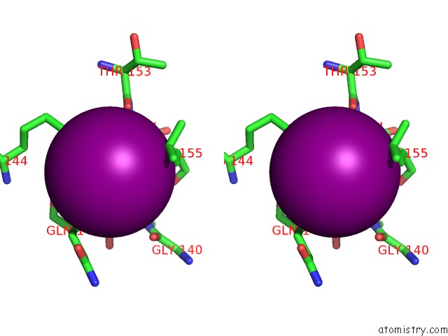
Iodine binding site 2 out of 4 in 2yd9
Go back to
Iodine binding site 2 out
of 4 in the Crystal Structure of the N-Terminal IG1-3 Module of Human Receptor Protein Tyrosine Phosphatase Sigma
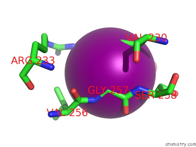
Mono view
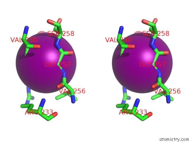
Stereo pair view
Mono view
Stereo pair view
A full contact list of Iodine with other atoms in the I binding
site number 2 of Crystal Structure of the N-Terminal IG1-3 Module of Human Receptor Protein Tyrosine Phosphatase Sigma within 5.0Å range:
|
Iodine binding site 3 out of 4 in 2yd9
Go back to
Iodine binding site 3 out
of 4 in the Crystal Structure of the N-Terminal IG1-3 Module of Human Receptor Protein Tyrosine Phosphatase Sigma
Mono view
Stereo pair view
Mono view
Stereo pair view
A full contact list of Iodine with other atoms in the I binding
site number 3 of Crystal Structure of the N-Terminal IG1-3 Module of Human Receptor Protein Tyrosine Phosphatase Sigma within 5.0Å range:
|
Iodine binding site 4 out of 4 in 2yd9
Go back to
Iodine binding site 4 out
of 4 in the Crystal Structure of the N-Terminal IG1-3 Module of Human Receptor Protein Tyrosine Phosphatase Sigma
Mono view
Stereo pair view
Mono view
Stereo pair view
A full contact list of Iodine with other atoms in the I binding
site number 4 of Crystal Structure of the N-Terminal IG1-3 Module of Human Receptor Protein Tyrosine Phosphatase Sigma within 5.0Å range:
|
Reference:
C.H.Coles,
Y.Shen,
A.P.Tenney,
C.Siebold,
G.C.Sutton,
W.Lu,
J.T.Gallagher,
E.Y.Jones,
J.G.Flanagan,
A.R.Aricescu.
Proteoglycan-Specific Molecular Switch For Rptp Sigma Clustering and Neuronal Extension. Science V. 332 484 2011.
ISSN: ISSN 0036-8075
PubMed: 21454754
DOI: 10.1126/SCIENCE.1200840
Page generated: Sun Aug 11 14:37:45 2024
ISSN: ISSN 0036-8075
PubMed: 21454754
DOI: 10.1126/SCIENCE.1200840
Last articles
Zn in 9J0NZn in 9J0O
Zn in 9J0P
Zn in 9FJX
Zn in 9EKB
Zn in 9C0F
Zn in 9CAH
Zn in 9CH0
Zn in 9CH3
Zn in 9CH1