Iodine »
PDB 2y6q-3cgp »
2yv7 »
Iodine in PDB 2yv7: Crystal Structure of the Clic Homolog From Drosophila Melanogaster
Protein crystallography data
The structure of Crystal Structure of the Clic Homolog From Drosophila Melanogaster, PDB code: 2yv7
was solved by
S.J.Harrop,
D.R.Littler,
P.M.G.Curmi,
with X-Ray Crystallography technique. A brief refinement statistics is given in the table below:
| Resolution Low / High (Å) | 32.00 / 1.70 |
| Space group | P 21 21 21 |
| Cell size a, b, c (Å), α, β, γ (°) | 39.393, 63.451, 114.122, 90.00, 90.00, 90.00 |
| R / Rfree (%) | 21.6 / 25.4 |
Other elements in 2yv7:
The structure of Crystal Structure of the Clic Homolog From Drosophila Melanogaster also contains other interesting chemical elements:
| Calcium | (Ca) | 1 atom |
Iodine Binding Sites:
The binding sites of Iodine atom in the Crystal Structure of the Clic Homolog From Drosophila Melanogaster
(pdb code 2yv7). This binding sites where shown within
5.0 Angstroms radius around Iodine atom.
In total only one binding site of Iodine was determined in the Crystal Structure of the Clic Homolog From Drosophila Melanogaster, PDB code: 2yv7:
In total only one binding site of Iodine was determined in the Crystal Structure of the Clic Homolog From Drosophila Melanogaster, PDB code: 2yv7:
Iodine binding site 1 out of 1 in 2yv7
Go back to
Iodine binding site 1 out
of 1 in the Crystal Structure of the Clic Homolog From Drosophila Melanogaster
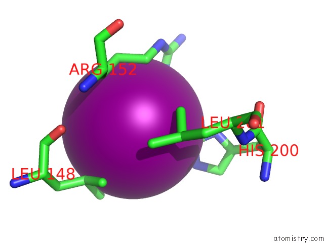
Mono view
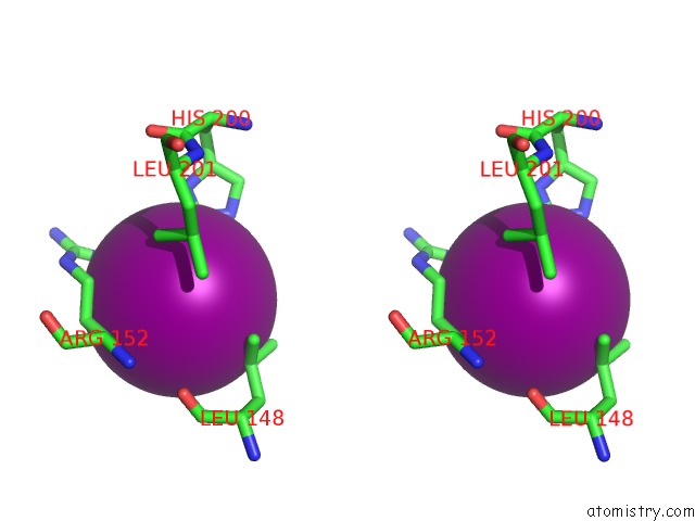
Stereo pair view
Mono view
Stereo pair view
A full contact list of Iodine with other atoms in the I binding
site number 1 of Crystal Structure of the Clic Homolog From Drosophila Melanogaster within 5.0Å range:
|
Reference:
D.R.Littler,
S.J.Harrop,
L.J.Brown,
G.J.Pankhurst,
A.V.Mynott,
P.Luciani,
R.A.Mandyam,
M.Mazzanti,
S.Tanda,
M.A.Berryman,
S.N.Breit,
P.M.G.Curmi.
Comparison of Vertebrate and Invertebrate Clic Proteins: the Crystal Structures of Caenorhabditis Elegans Exc-4 and Drosophila Melanogaster Dmclic Proteins V. 71 364 2007.
ISSN: ISSN 0887-3585
PubMed: 17985355
DOI: 10.1002/PROT.21704
Page generated: Sun Aug 11 14:39:16 2024
ISSN: ISSN 0887-3585
PubMed: 17985355
DOI: 10.1002/PROT.21704
Last articles
Zn in 9J0NZn in 9J0O
Zn in 9J0P
Zn in 9FJX
Zn in 9EKB
Zn in 9C0F
Zn in 9CAH
Zn in 9CH0
Zn in 9CH3
Zn in 9CH1