Iodine »
PDB 2y6q-3cgp »
2zxv »
Iodine in PDB 2zxv: Crystal Structure of Putative Acetyltransferase From T. Thermophilus HB8
Enzymatic activity of Crystal Structure of Putative Acetyltransferase From T. Thermophilus HB8
All present enzymatic activity of Crystal Structure of Putative Acetyltransferase From T. Thermophilus HB8:
2.3.1.128;
2.3.1.128;
Protein crystallography data
The structure of Crystal Structure of Putative Acetyltransferase From T. Thermophilus HB8, PDB code: 2zxv
was solved by
K.Murayama,
M.Kato-Murayama,
T.Terada,
S.Kuramitsu,
M.Shirouzu,
S.Yokoyama,
Riken Structural Genomics/Proteomics Initiative(Rsgi),
with X-Ray Crystallography technique. A brief refinement statistics is given in the table below:
| Resolution Low / High (Å) | 30.13 / 2.30 |
| Space group | I 4 |
| Cell size a, b, c (Å), α, β, γ (°) | 127.800, 127.800, 122.100, 90.00, 90.00, 90.00 |
| R / Rfree (%) | 22.8 / 26.6 |
Iodine Binding Sites:
The binding sites of Iodine atom in the Crystal Structure of Putative Acetyltransferase From T. Thermophilus HB8
(pdb code 2zxv). This binding sites where shown within
5.0 Angstroms radius around Iodine atom.
In total 4 binding sites of Iodine where determined in the Crystal Structure of Putative Acetyltransferase From T. Thermophilus HB8, PDB code: 2zxv:
Jump to Iodine binding site number: 1; 2; 3; 4;
In total 4 binding sites of Iodine where determined in the Crystal Structure of Putative Acetyltransferase From T. Thermophilus HB8, PDB code: 2zxv:
Jump to Iodine binding site number: 1; 2; 3; 4;
Iodine binding site 1 out of 4 in 2zxv
Go back to
Iodine binding site 1 out
of 4 in the Crystal Structure of Putative Acetyltransferase From T. Thermophilus HB8

Mono view
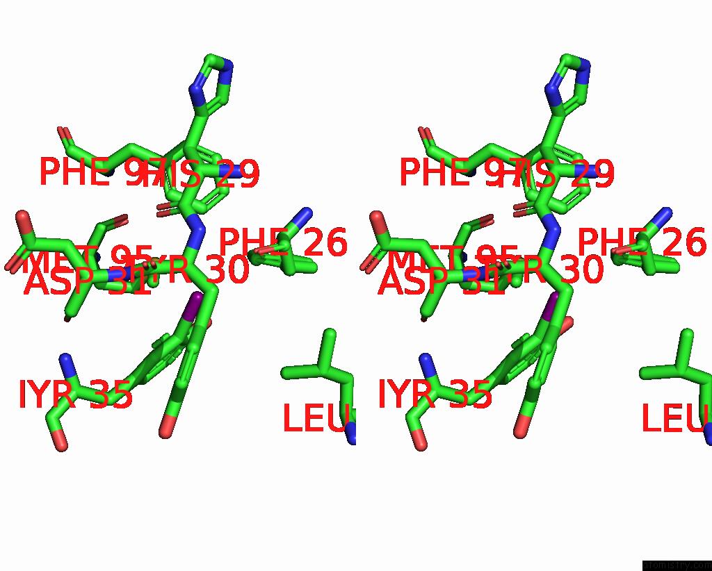
Stereo pair view
Mono view
Stereo pair view
A full contact list of Iodine with other atoms in the I binding
site number 1 of Crystal Structure of Putative Acetyltransferase From T. Thermophilus HB8 within 5.0Å range:
|
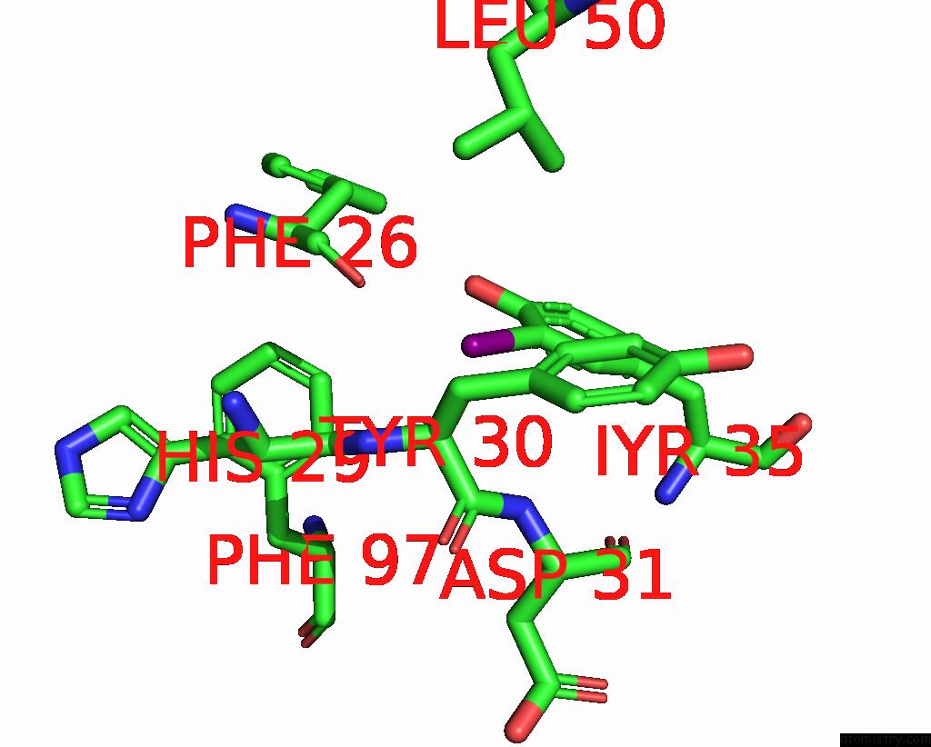
Iodine binding site 2 out of 4 in 2zxv
Go back to
Iodine binding site 2 out
of 4 in the Crystal Structure of Putative Acetyltransferase From T. Thermophilus HB8

Mono view

Stereo pair view
Mono view
Stereo pair view
A full contact list of Iodine with other atoms in the I binding
site number 2 of Crystal Structure of Putative Acetyltransferase From T. Thermophilus HB8 within 5.0Å range:
|
Iodine binding site 3 out of 4 in 2zxv
Go back to
Iodine binding site 3 out
of 4 in the Crystal Structure of Putative Acetyltransferase From T. Thermophilus HB8
Mono view
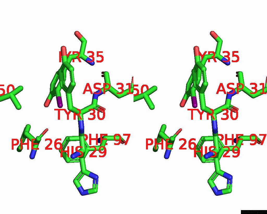
Stereo pair view
Mono view
Stereo pair view
A full contact list of Iodine with other atoms in the I binding
site number 3 of Crystal Structure of Putative Acetyltransferase From T. Thermophilus HB8 within 5.0Å range:
|
Iodine binding site 4 out of 4 in 2zxv
Go back to
Iodine binding site 4 out
of 4 in the Crystal Structure of Putative Acetyltransferase From T. Thermophilus HB8
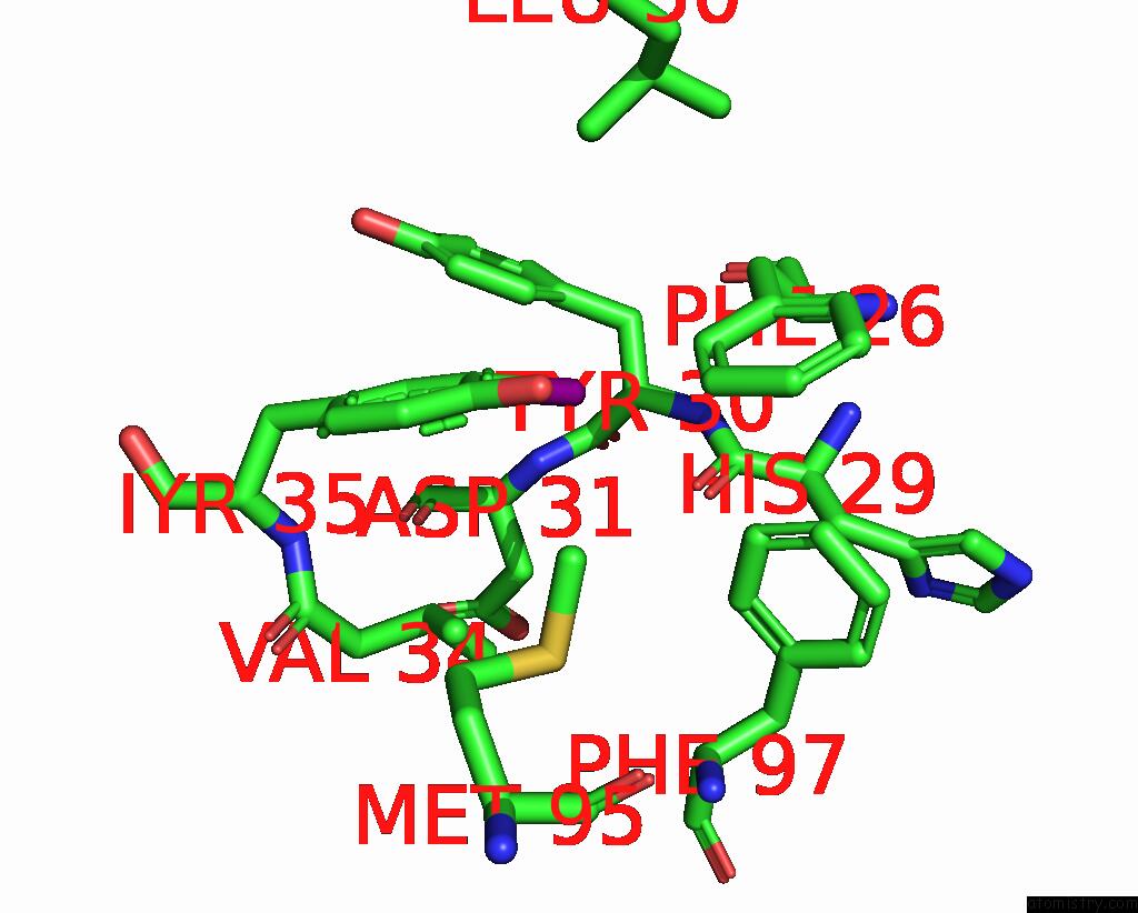
Mono view

Stereo pair view
Mono view
Stereo pair view
A full contact list of Iodine with other atoms in the I binding
site number 4 of Crystal Structure of Putative Acetyltransferase From T. Thermophilus HB8 within 5.0Å range:
|
Reference:
K.Sakamoto,
K.Murayama,
K.Oki,
F.Iraha,
M.Kato-Murayama,
M.Takahashi,
K.Ohtake,
T.Kobayashi,
S.Kuramitsu,
M.Shirouzu,
S.Yokoyama.
Genetic Encoding of 3-Iodo-L-Tyrosine in Escherichia Coli For Single-Wavelength Anomalous Dispersion Phasing in Protein Crystallography Structure V. 17 335 2009.
ISSN: ISSN 0969-2126
PubMed: 19278648
DOI: 10.1016/J.STR.2009.01.008
Page generated: Sun Aug 11 14:39:52 2024
ISSN: ISSN 0969-2126
PubMed: 19278648
DOI: 10.1016/J.STR.2009.01.008
Last articles
Zn in 9J0NZn in 9J0O
Zn in 9J0P
Zn in 9FJX
Zn in 9EKB
Zn in 9C0F
Zn in 9CAH
Zn in 9CH0
Zn in 9CH3
Zn in 9CH1