Iodine »
PDB 2y6q-3cgp »
3apb »
Iodine in PDB 3apb: Crystal Structure of the Galectin-8 N-Terminal Carbohydrate Recognition Domain in Complex with Iodide
Protein crystallography data
The structure of Crystal Structure of the Galectin-8 N-Terminal Carbohydrate Recognition Domain in Complex with Iodide, PDB code: 3apb
was solved by
T.Matsuzaka,
H.Ideo,
K.Yamashita,
T.Nonaka,
with X-Ray Crystallography technique. A brief refinement statistics is given in the table below:
| Resolution Low / High (Å) | 38.95 / 1.95 |
| Space group | P 21 21 21 |
| Cell size a, b, c (Å), α, β, γ (°) | 65.591, 75.869, 77.901, 90.00, 90.00, 90.00 |
| R / Rfree (%) | 20.2 / 24.2 |
Iodine Binding Sites:
Pages:
>>> Page 1 <<< Page 2, Binding sites: 11 - 20; Page 3, Binding sites: 21 - 29;Binding sites:
The binding sites of Iodine atom in the Crystal Structure of the Galectin-8 N-Terminal Carbohydrate Recognition Domain in Complex with Iodide (pdb code 3apb). This binding sites where shown within 5.0 Angstroms radius around Iodine atom.In total 29 binding sites of Iodine where determined in the Crystal Structure of the Galectin-8 N-Terminal Carbohydrate Recognition Domain in Complex with Iodide, PDB code: 3apb:
Jump to Iodine binding site number: 1; 2; 3; 4; 5; 6; 7; 8; 9; 10;
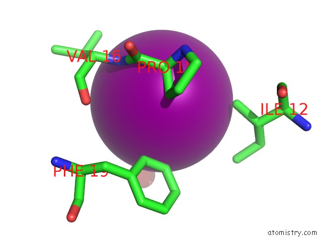
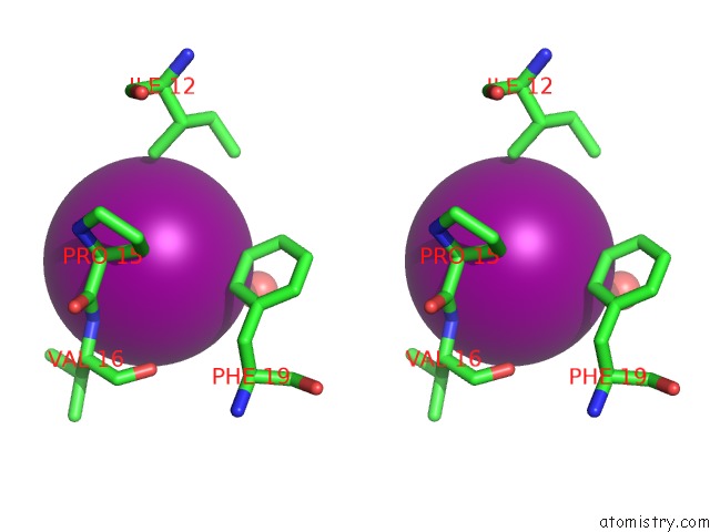
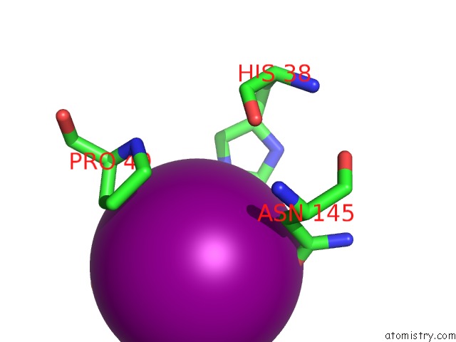
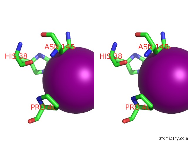
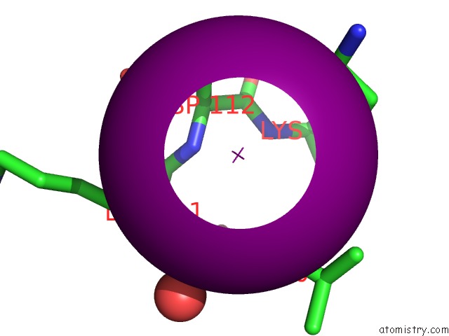
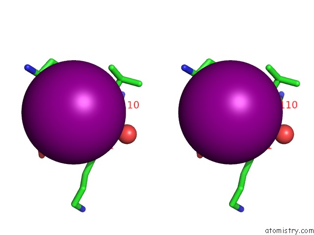
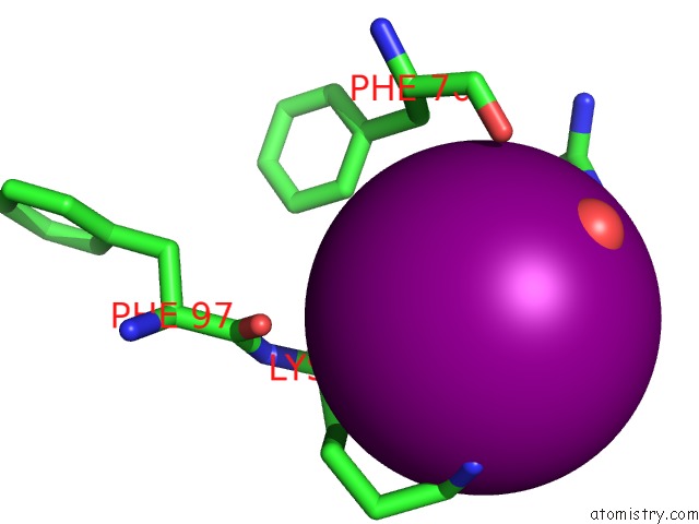
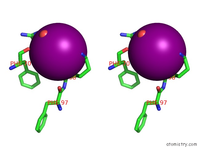
Iodine binding site 1 out of 29 in 3apb
Go back to
Iodine binding site 1 out
of 29 in the Crystal Structure of the Galectin-8 N-Terminal Carbohydrate Recognition Domain in Complex with Iodide
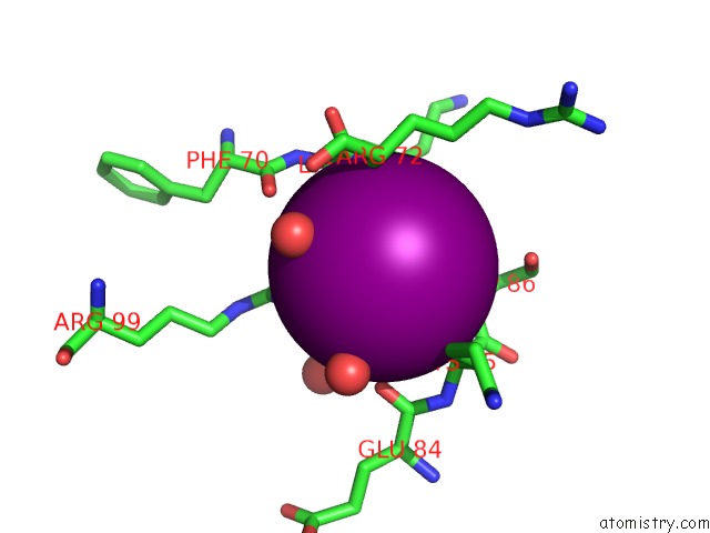
Mono view
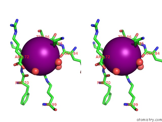
Stereo pair view
Mono view
Stereo pair view
A full contact list of Iodine with other atoms in the I binding
site number 1 of Crystal Structure of the Galectin-8 N-Terminal Carbohydrate Recognition Domain in Complex with Iodide within 5.0Å range:
|
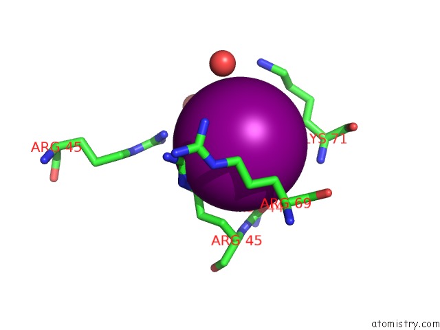
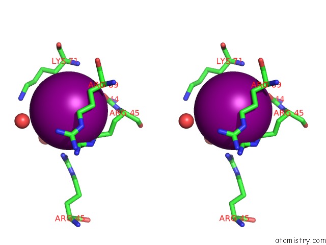
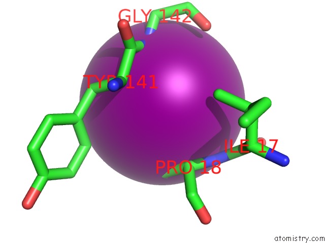
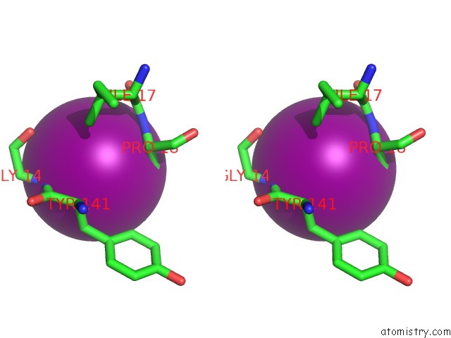
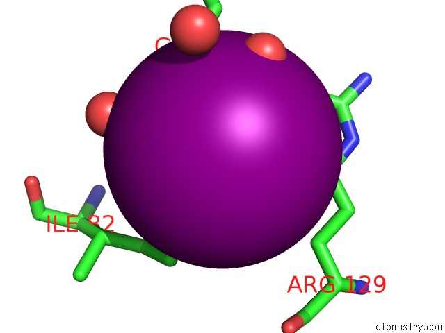
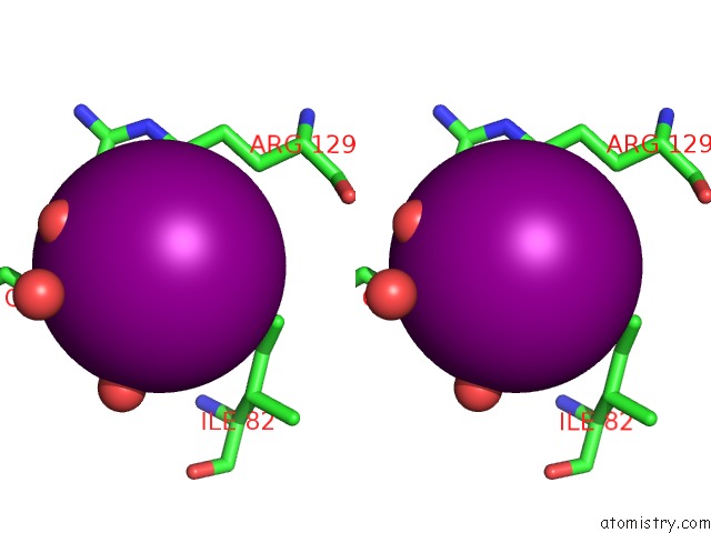
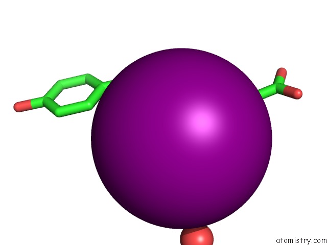
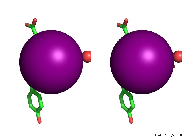
Iodine binding site 2 out of 29 in 3apb
Go back to
Iodine binding site 2 out
of 29 in the Crystal Structure of the Galectin-8 N-Terminal Carbohydrate Recognition Domain in Complex with Iodide
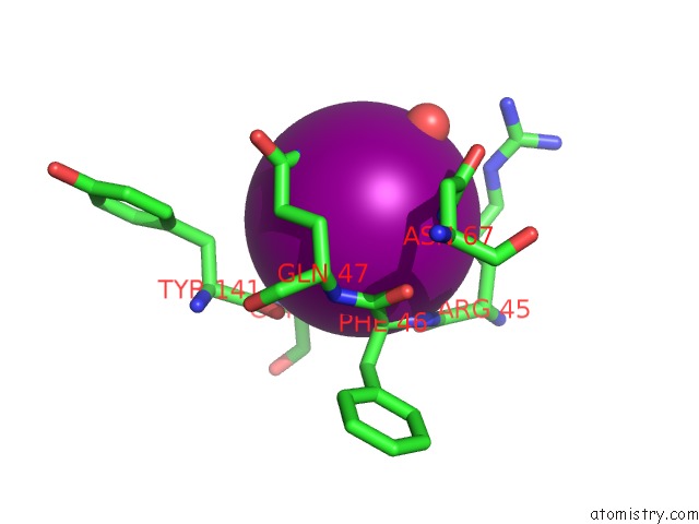
Mono view
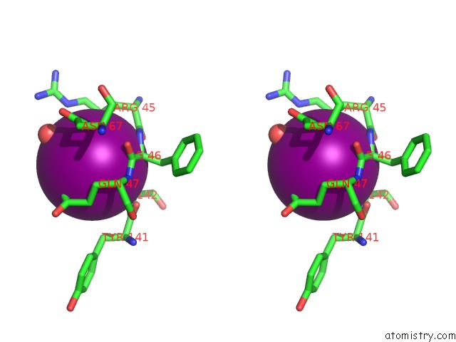
Stereo pair view
Mono view
Stereo pair view
A full contact list of Iodine with other atoms in the I binding
site number 2 of Crystal Structure of the Galectin-8 N-Terminal Carbohydrate Recognition Domain in Complex with Iodide within 5.0Å range:
|
Iodine binding site 3 out of 29 in 3apb
Go back to
Iodine binding site 3 out
of 29 in the Crystal Structure of the Galectin-8 N-Terminal Carbohydrate Recognition Domain in Complex with Iodide
Mono view
Stereo pair view
Mono view
Stereo pair view
A full contact list of Iodine with other atoms in the I binding
site number 3 of Crystal Structure of the Galectin-8 N-Terminal Carbohydrate Recognition Domain in Complex with Iodide within 5.0Å range:
|
Iodine binding site 4 out of 29 in 3apb
Go back to
Iodine binding site 4 out
of 29 in the Crystal Structure of the Galectin-8 N-Terminal Carbohydrate Recognition Domain in Complex with Iodide
Mono view
Stereo pair view
Mono view
Stereo pair view
A full contact list of Iodine with other atoms in the I binding
site number 4 of Crystal Structure of the Galectin-8 N-Terminal Carbohydrate Recognition Domain in Complex with Iodide within 5.0Å range:
|
Iodine binding site 5 out of 29 in 3apb
Go back to
Iodine binding site 5 out
of 29 in the Crystal Structure of the Galectin-8 N-Terminal Carbohydrate Recognition Domain in Complex with Iodide
Mono view
Stereo pair view
Mono view
Stereo pair view
A full contact list of Iodine with other atoms in the I binding
site number 5 of Crystal Structure of the Galectin-8 N-Terminal Carbohydrate Recognition Domain in Complex with Iodide within 5.0Å range:
|
Iodine binding site 6 out of 29 in 3apb
Go back to
Iodine binding site 6 out
of 29 in the Crystal Structure of the Galectin-8 N-Terminal Carbohydrate Recognition Domain in Complex with Iodide
Mono view
Stereo pair view
Mono view
Stereo pair view
A full contact list of Iodine with other atoms in the I binding
site number 6 of Crystal Structure of the Galectin-8 N-Terminal Carbohydrate Recognition Domain in Complex with Iodide within 5.0Å range:
|
Iodine binding site 7 out of 29 in 3apb
Go back to
Iodine binding site 7 out
of 29 in the Crystal Structure of the Galectin-8 N-Terminal Carbohydrate Recognition Domain in Complex with Iodide
Mono view
Stereo pair view
Mono view
Stereo pair view
A full contact list of Iodine with other atoms in the I binding
site number 7 of Crystal Structure of the Galectin-8 N-Terminal Carbohydrate Recognition Domain in Complex with Iodide within 5.0Å range:
|
Iodine binding site 8 out of 29 in 3apb
Go back to
Iodine binding site 8 out
of 29 in the Crystal Structure of the Galectin-8 N-Terminal Carbohydrate Recognition Domain in Complex with Iodide
Mono view
Stereo pair view
Mono view
Stereo pair view
A full contact list of Iodine with other atoms in the I binding
site number 8 of Crystal Structure of the Galectin-8 N-Terminal Carbohydrate Recognition Domain in Complex with Iodide within 5.0Å range:
|
Iodine binding site 9 out of 29 in 3apb
Go back to
Iodine binding site 9 out
of 29 in the Crystal Structure of the Galectin-8 N-Terminal Carbohydrate Recognition Domain in Complex with Iodide
Mono view
Stereo pair view
Mono view
Stereo pair view
A full contact list of Iodine with other atoms in the I binding
site number 9 of Crystal Structure of the Galectin-8 N-Terminal Carbohydrate Recognition Domain in Complex with Iodide within 5.0Å range:
|
Iodine binding site 10 out of 29 in 3apb
Go back to
Iodine binding site 10 out
of 29 in the Crystal Structure of the Galectin-8 N-Terminal Carbohydrate Recognition Domain in Complex with Iodide
Mono view
Stereo pair view
Mono view
Stereo pair view
A full contact list of Iodine with other atoms in the I binding
site number 10 of Crystal Structure of the Galectin-8 N-Terminal Carbohydrate Recognition Domain in Complex with Iodide within 5.0Å range:
|
Reference:
H.Ideo,
T.Matsuzaka,
T.Nonaka,
A.Seko,
K.Yamashita.
Galectin-8-N-Domain Recognition Mechanism For Sialylated and Sulfated Glycans To Be Published.
Page generated: Fri Aug 8 13:53:32 2025
Last articles
I in 4DUSI in 4DHG
I in 4DH6
I in 4DNY
I in 4CB6
I in 4DCH
I in 4BH5
I in 4BVA
I in 4D85
I in 4CJD