Iodine »
PDB 2y6q-3cgp »
3b56 »
Iodine in PDB 3b56: Crystal Structure of Transthyretin in Complex with 3,5- Diiodosalicylic Acid
Protein crystallography data
The structure of Crystal Structure of Transthyretin in Complex with 3,5- Diiodosalicylic Acid, PDB code: 3b56
was solved by
L.Gales,
A.M.Damas,
with X-Ray Crystallography technique. A brief refinement statistics is given in the table below:
| Resolution Low / High (Å) | 27.34 / 1.55 |
| Space group | P 21 21 2 |
| Cell size a, b, c (Å), α, β, γ (°) | 42.539, 85.757, 64.346, 90.00, 90.00, 90.00 |
| R / Rfree (%) | 18.9 / 21.4 |
Iodine Binding Sites:
The binding sites of Iodine atom in the Crystal Structure of Transthyretin in Complex with 3,5- Diiodosalicylic Acid
(pdb code 3b56). This binding sites where shown within
5.0 Angstroms radius around Iodine atom.
In total 4 binding sites of Iodine where determined in the Crystal Structure of Transthyretin in Complex with 3,5- Diiodosalicylic Acid, PDB code: 3b56:
Jump to Iodine binding site number: 1; 2; 3; 4;
In total 4 binding sites of Iodine where determined in the Crystal Structure of Transthyretin in Complex with 3,5- Diiodosalicylic Acid, PDB code: 3b56:
Jump to Iodine binding site number: 1; 2; 3; 4;
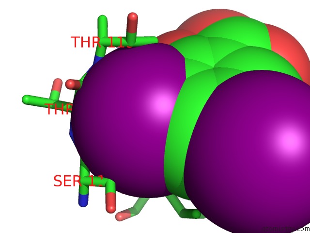
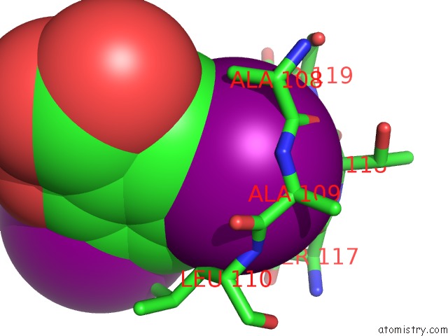
Iodine binding site 1 out of 4 in 3b56
Go back to
Iodine binding site 1 out
of 4 in the Crystal Structure of Transthyretin in Complex with 3,5- Diiodosalicylic Acid
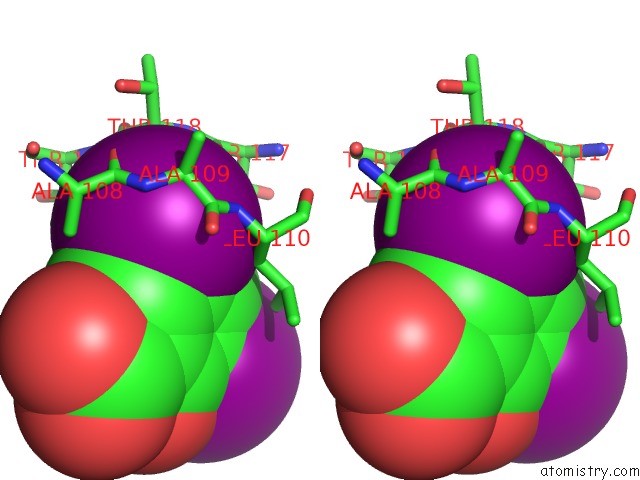
Mono view
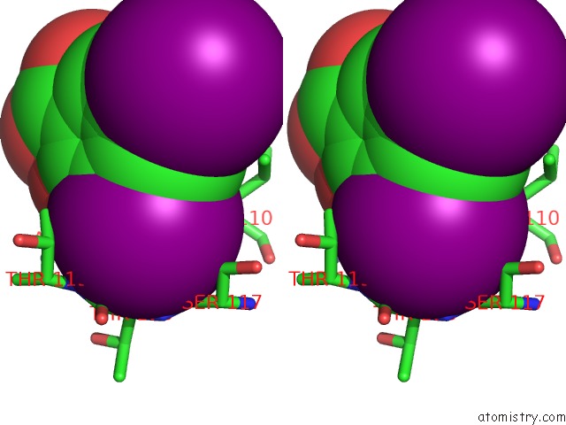
Stereo pair view
Mono view
Stereo pair view
A full contact list of Iodine with other atoms in the I binding
site number 1 of Crystal Structure of Transthyretin in Complex with 3,5- Diiodosalicylic Acid within 5.0Å range:
|
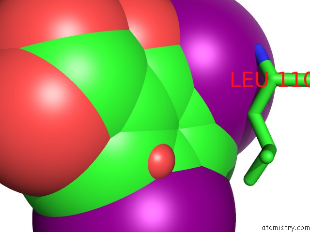
Iodine binding site 2 out of 4 in 3b56
Go back to
Iodine binding site 2 out
of 4 in the Crystal Structure of Transthyretin in Complex with 3,5- Diiodosalicylic Acid
Mono view
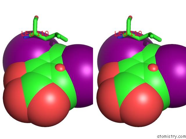
Stereo pair view
Mono view
Stereo pair view
A full contact list of Iodine with other atoms in the I binding
site number 2 of Crystal Structure of Transthyretin in Complex with 3,5- Diiodosalicylic Acid within 5.0Å range:
|
Iodine binding site 3 out of 4 in 3b56
Go back to
Iodine binding site 3 out
of 4 in the Crystal Structure of Transthyretin in Complex with 3,5- Diiodosalicylic Acid
Mono view

Stereo pair view
Mono view
Stereo pair view
A full contact list of Iodine with other atoms in the I binding
site number 3 of Crystal Structure of Transthyretin in Complex with 3,5- Diiodosalicylic Acid within 5.0Å range:
|
Iodine binding site 4 out of 4 in 3b56
Go back to
Iodine binding site 4 out
of 4 in the Crystal Structure of Transthyretin in Complex with 3,5- Diiodosalicylic Acid
Mono view
Stereo pair view
Mono view
Stereo pair view
A full contact list of Iodine with other atoms in the I binding
site number 4 of Crystal Structure of Transthyretin in Complex with 3,5- Diiodosalicylic Acid within 5.0Å range:
|
Reference:
L.Gales,
M.R.Almeida,
G.Arsequell,
G.Valencia,
M.J.Saraiva,
A.M.Damas.
Iodination of Salicylic Acid Improves Its Binding to Transthyretin Biochim.Biophys.Acta V.1784 512 2008.
ISSN: ISSN 0006-3002
PubMed: 18155178
DOI: 10.1016/J.BBAPAP.2007.11.014
Page generated: Fri Aug 8 13:57:44 2025
ISSN: ISSN 0006-3002
PubMed: 18155178
DOI: 10.1016/J.BBAPAP.2007.11.014
Last articles
I in 4DUSI in 4DHG
I in 4DH6
I in 4DNY
I in 4CB6
I in 4DCH
I in 4BH5
I in 4BVA
I in 4D85
I in 4CJD