Iodine »
PDB 3cjq-3fnl »
3d7f »
Iodine in PDB 3d7f: A High Resolution Crystal Structure of Human Glutamate Carboxypeptidase II (Gcpii) in A Complex with Dcit, A Urea-Based Inhibitor
Enzymatic activity of A High Resolution Crystal Structure of Human Glutamate Carboxypeptidase II (Gcpii) in A Complex with Dcit, A Urea-Based Inhibitor
All present enzymatic activity of A High Resolution Crystal Structure of Human Glutamate Carboxypeptidase II (Gcpii) in A Complex with Dcit, A Urea-Based Inhibitor:
3.4.17.21;
3.4.17.21;
Protein crystallography data
The structure of A High Resolution Crystal Structure of Human Glutamate Carboxypeptidase II (Gcpii) in A Complex with Dcit, A Urea-Based Inhibitor, PDB code: 3d7f
was solved by
J.Lubkowski,
C.Barinka,
with X-Ray Crystallography technique. A brief refinement statistics is given in the table below:
| Resolution Low / High (Å) | 15.00 / 1.54 |
| Space group | I 2 2 2 |
| Cell size a, b, c (Å), α, β, γ (°) | 101.434, 130.059, 158.543, 90.00, 90.00, 90.00 |
| R / Rfree (%) | 17.7 / 19.5 |
Other elements in 3d7f:
The structure of A High Resolution Crystal Structure of Human Glutamate Carboxypeptidase II (Gcpii) in A Complex with Dcit, A Urea-Based Inhibitor also contains other interesting chemical elements:
| Chlorine | (Cl) | 1 atom |
| Calcium | (Ca) | 1 atom |
| Zinc | (Zn) | 2 atoms |
Iodine Binding Sites:
The binding sites of Iodine atom in the A High Resolution Crystal Structure of Human Glutamate Carboxypeptidase II (Gcpii) in A Complex with Dcit, A Urea-Based Inhibitor
(pdb code 3d7f). This binding sites where shown within
5.0 Angstroms radius around Iodine atom.
In total only one binding site of Iodine was determined in the A High Resolution Crystal Structure of Human Glutamate Carboxypeptidase II (Gcpii) in A Complex with Dcit, A Urea-Based Inhibitor, PDB code: 3d7f:
In total only one binding site of Iodine was determined in the A High Resolution Crystal Structure of Human Glutamate Carboxypeptidase II (Gcpii) in A Complex with Dcit, A Urea-Based Inhibitor, PDB code: 3d7f:
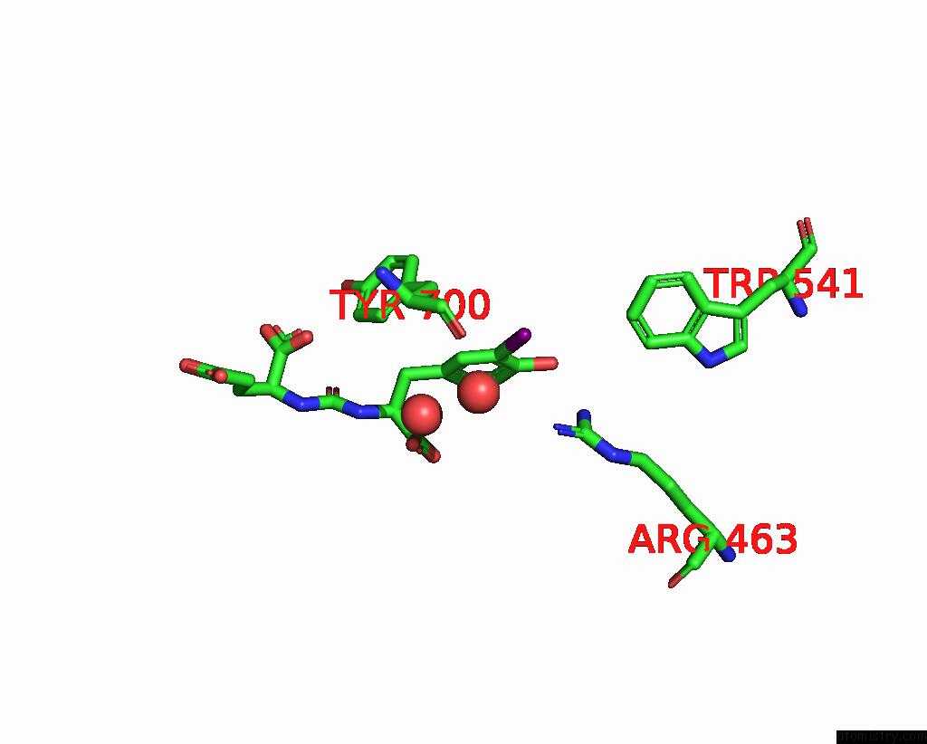
Iodine binding site 1 out of 1 in 3d7f
Go back to
Iodine binding site 1 out
of 1 in the A High Resolution Crystal Structure of Human Glutamate Carboxypeptidase II (Gcpii) in A Complex with Dcit, A Urea-Based Inhibitor
Mono view

Stereo pair view
Mono view
Stereo pair view
A full contact list of Iodine with other atoms in the I binding
site number 1 of A High Resolution Crystal Structure of Human Glutamate Carboxypeptidase II (Gcpii) in A Complex with Dcit, A Urea-Based Inhibitor within 5.0Å range:
|
Reference:
C.Barinka,
Y.Byun,
C.L.Dusich,
S.R.Banerjee,
Y.Chen,
M.Castanares,
A.P.Kozikowski,
R.C.Mease,
M.G.Pomper,
J.Lubkowski.
Interactions Between Human Glutamate Carboxypeptidase II and Urea-Based Inhibitors: Structural Characterization J.Med.Chem. V. 51 7737 2008.
ISSN: ISSN 0022-2623
PubMed: 19053759
DOI: 10.1021/JM800765E
Page generated: Fri Aug 8 14:04:31 2025
ISSN: ISSN 0022-2623
PubMed: 19053759
DOI: 10.1021/JM800765E
Last articles
I in 4LDVI in 4LBR
I in 4LB3
I in 4LAZ
I in 4KSZ
I in 4L6C
I in 4K1C
I in 4KQV
I in 4KRW
I in 4KRL