Iodine »
PDB 3cjq-3fnl »
3da8 »
Iodine in PDB 3da8: Crystal Structure of Purn From Mycobacterium Tuberculosis
Enzymatic activity of Crystal Structure of Purn From Mycobacterium Tuberculosis
All present enzymatic activity of Crystal Structure of Purn From Mycobacterium Tuberculosis:
2.1.2.2;
2.1.2.2;
Protein crystallography data
The structure of Crystal Structure of Purn From Mycobacterium Tuberculosis, PDB code: 3da8
was solved by
Z.Zhang,
C.J.Squire,
E.N.Baker,
with X-Ray Crystallography technique. A brief refinement statistics is given in the table below:
| Resolution Low / High (Å) | 36.69 / 1.30 |
| Space group | P 21 21 21 |
| Cell size a, b, c (Å), α, β, γ (°) | 73.273, 73.366, 77.246, 90.00, 90.00, 90.00 |
| R / Rfree (%) | 16.4 / 18.6 |
Other elements in 3da8:
The structure of Crystal Structure of Purn From Mycobacterium Tuberculosis also contains other interesting chemical elements:
| Magnesium | (Mg) | 1 atom |
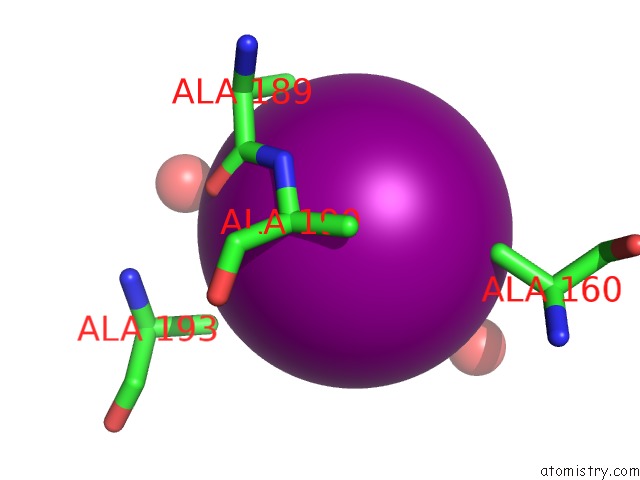
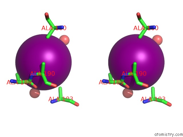
Iodine Binding Sites:
The binding sites of Iodine atom in the Crystal Structure of Purn From Mycobacterium Tuberculosis
(pdb code 3da8). This binding sites where shown within
5.0 Angstroms radius around Iodine atom.
In total 9 binding sites of Iodine where determined in the Crystal Structure of Purn From Mycobacterium Tuberculosis, PDB code: 3da8:
Jump to Iodine binding site number: 1; 2; 3; 4; 5; 6; 7; 8; 9;
In total 9 binding sites of Iodine where determined in the Crystal Structure of Purn From Mycobacterium Tuberculosis, PDB code: 3da8:
Jump to Iodine binding site number: 1; 2; 3; 4; 5; 6; 7; 8; 9;
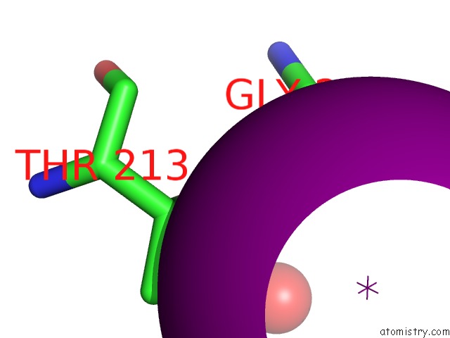
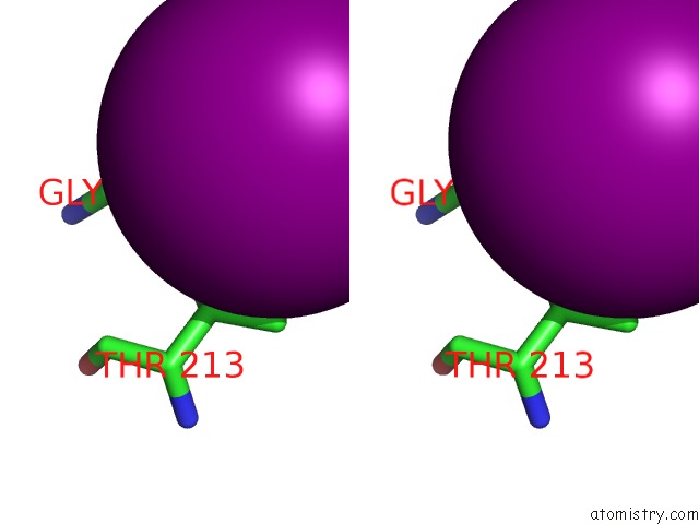
Iodine binding site 1 out of 9 in 3da8
Go back to
Iodine binding site 1 out
of 9 in the Crystal Structure of Purn From Mycobacterium Tuberculosis
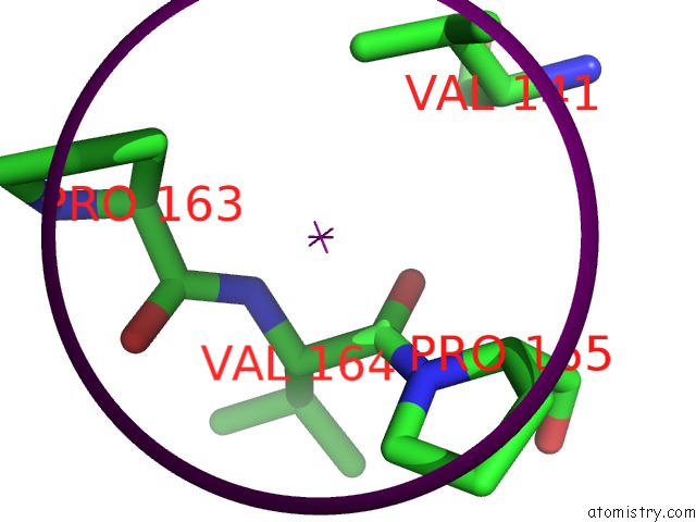
Mono view
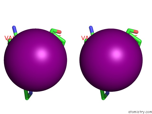
Stereo pair view
Mono view
Stereo pair view
A full contact list of Iodine with other atoms in the I binding
site number 1 of Crystal Structure of Purn From Mycobacterium Tuberculosis within 5.0Å range:
|
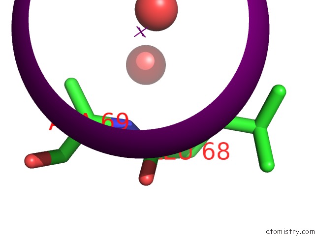
Iodine binding site 2 out of 9 in 3da8
Go back to
Iodine binding site 2 out
of 9 in the Crystal Structure of Purn From Mycobacterium Tuberculosis
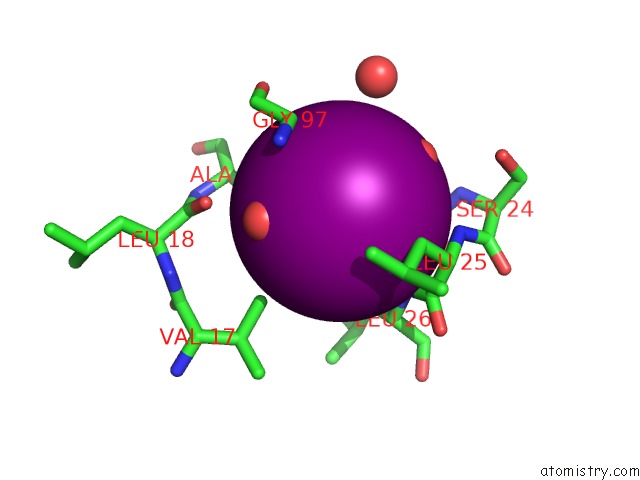
Mono view
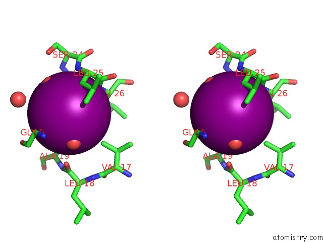
Stereo pair view
Mono view
Stereo pair view
A full contact list of Iodine with other atoms in the I binding
site number 2 of Crystal Structure of Purn From Mycobacterium Tuberculosis within 5.0Å range:
|
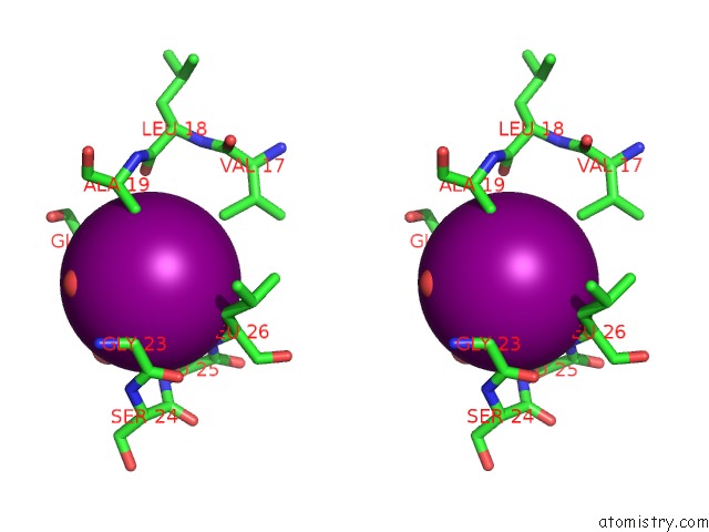
Iodine binding site 3 out of 9 in 3da8
Go back to
Iodine binding site 3 out
of 9 in the Crystal Structure of Purn From Mycobacterium Tuberculosis

Mono view

Stereo pair view
Mono view
Stereo pair view
A full contact list of Iodine with other atoms in the I binding
site number 3 of Crystal Structure of Purn From Mycobacterium Tuberculosis within 5.0Å range:
|
Iodine binding site 4 out of 9 in 3da8
Go back to
Iodine binding site 4 out
of 9 in the Crystal Structure of Purn From Mycobacterium Tuberculosis
Mono view
Stereo pair view
Mono view
Stereo pair view
A full contact list of Iodine with other atoms in the I binding
site number 4 of Crystal Structure of Purn From Mycobacterium Tuberculosis within 5.0Å range:
|
Iodine binding site 5 out of 9 in 3da8
Go back to
Iodine binding site 5 out
of 9 in the Crystal Structure of Purn From Mycobacterium Tuberculosis
Mono view

Stereo pair view
Mono view
Stereo pair view
A full contact list of Iodine with other atoms in the I binding
site number 5 of Crystal Structure of Purn From Mycobacterium Tuberculosis within 5.0Å range:
|
Iodine binding site 6 out of 9 in 3da8
Go back to
Iodine binding site 6 out
of 9 in the Crystal Structure of Purn From Mycobacterium Tuberculosis

Mono view

Stereo pair view
Mono view
Stereo pair view
A full contact list of Iodine with other atoms in the I binding
site number 6 of Crystal Structure of Purn From Mycobacterium Tuberculosis within 5.0Å range:
|
Iodine binding site 7 out of 9 in 3da8
Go back to
Iodine binding site 7 out
of 9 in the Crystal Structure of Purn From Mycobacterium Tuberculosis

Mono view
Stereo pair view
Mono view
Stereo pair view
A full contact list of Iodine with other atoms in the I binding
site number 7 of Crystal Structure of Purn From Mycobacterium Tuberculosis within 5.0Å range:
|
Iodine binding site 8 out of 9 in 3da8
Go back to
Iodine binding site 8 out
of 9 in the Crystal Structure of Purn From Mycobacterium Tuberculosis
Mono view
Stereo pair view
Mono view
Stereo pair view
A full contact list of Iodine with other atoms in the I binding
site number 8 of Crystal Structure of Purn From Mycobacterium Tuberculosis within 5.0Å range:
|
Iodine binding site 9 out of 9 in 3da8
Go back to
Iodine binding site 9 out
of 9 in the Crystal Structure of Purn From Mycobacterium Tuberculosis

Mono view

Stereo pair view
Mono view
Stereo pair view
A full contact list of Iodine with other atoms in the I binding
site number 9 of Crystal Structure of Purn From Mycobacterium Tuberculosis within 5.0Å range:
|
Reference:
Z.Zhang,
T.T.Caradoc-Davies,
J.M.Dickson,
E.N.Baker,
C.J.Squire.
Structures of Glycinamide Ribonucleotide Transformylase (Purn) From Mycobacterium Tuberculosis Reveal A Novel Dimer with Relevance to Drug Discovery. J.Mol.Biol. V. 389 722 2009.
ISSN: ISSN 0022-2836
PubMed: 19394344
DOI: 10.1016/J.JMB.2009.04.044
Page generated: Sun Aug 11 14:58:50 2024
ISSN: ISSN 0022-2836
PubMed: 19394344
DOI: 10.1016/J.JMB.2009.04.044
Last articles
Zn in 9J0NZn in 9J0O
Zn in 9J0P
Zn in 9FJX
Zn in 9EKB
Zn in 9C0F
Zn in 9CAH
Zn in 9CH0
Zn in 9CH3
Zn in 9CH1