Iodine »
PDB 3cjq-3fnl »
3dhx »
Iodine in PDB 3dhx: Crystal Structure of Isolated C2 Domain of the Methionine Uptake Transporter
Protein crystallography data
The structure of Crystal Structure of Isolated C2 Domain of the Methionine Uptake Transporter, PDB code: 3dhx
was solved by
E.Johnson,
J.T.Kaiser,
A.T.Lee,
D.C.Rees,
with X-Ray Crystallography technique. A brief refinement statistics is given in the table below:
| Resolution Low / High (Å) | 44.11 / 2.10 |
| Space group | P 31 2 1 |
| Cell size a, b, c (Å), α, β, γ (°) | 53.277, 53.277, 150.495, 90.00, 90.00, 120.00 |
| R / Rfree (%) | 19.1 / 23.8 |
Iodine Binding Sites:
Pages:
>>> Page 1 <<< Page 2, Binding sites: 11 - 12;Binding sites:
The binding sites of Iodine atom in the Crystal Structure of Isolated C2 Domain of the Methionine Uptake Transporter (pdb code 3dhx). This binding sites where shown within 5.0 Angstroms radius around Iodine atom.In total 12 binding sites of Iodine where determined in the Crystal Structure of Isolated C2 Domain of the Methionine Uptake Transporter, PDB code: 3dhx:
Jump to Iodine binding site number: 1; 2; 3; 4; 5; 6; 7; 8; 9; 10;
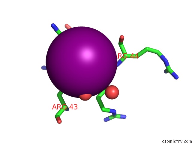
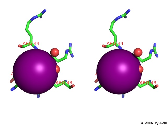
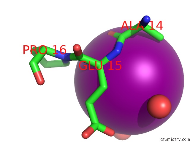
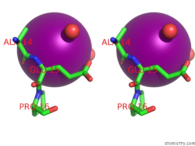
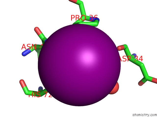
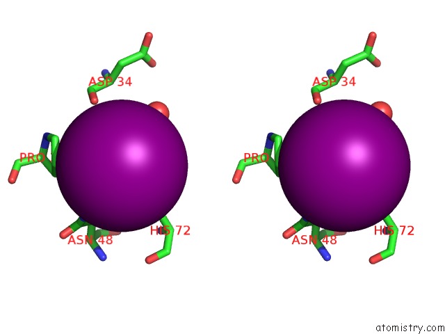
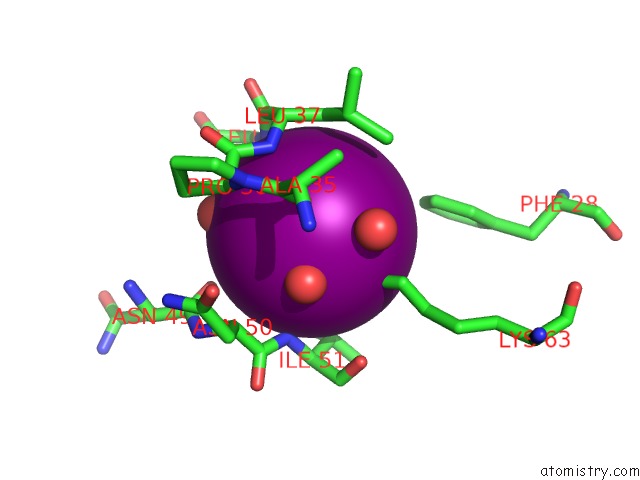
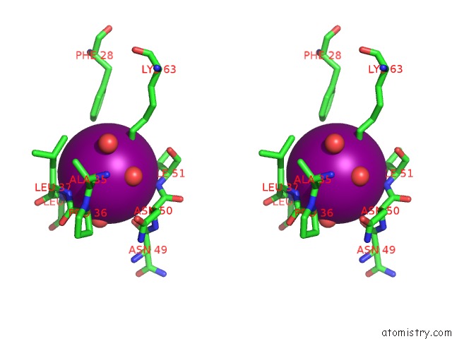
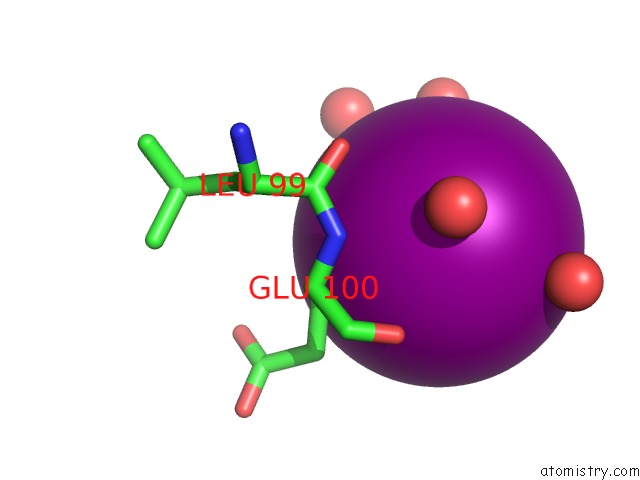
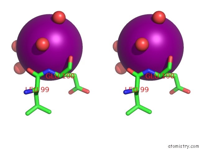
Iodine binding site 1 out of 12 in 3dhx
Go back to
Iodine binding site 1 out
of 12 in the Crystal Structure of Isolated C2 Domain of the Methionine Uptake Transporter
Mono view
Stereo pair view
Mono view
Stereo pair view
A full contact list of Iodine with other atoms in the I binding
site number 1 of Crystal Structure of Isolated C2 Domain of the Methionine Uptake Transporter within 5.0Å range:
|
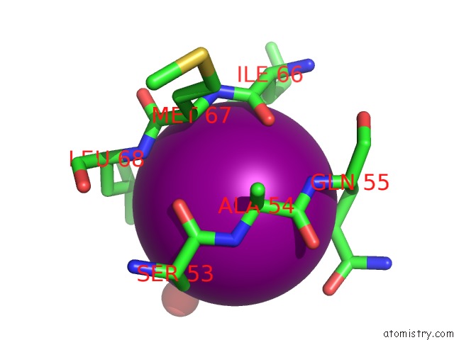
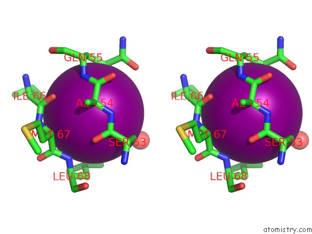
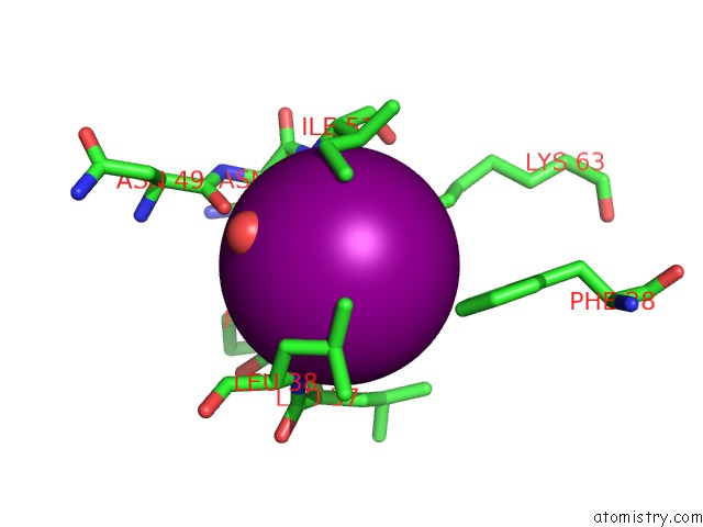
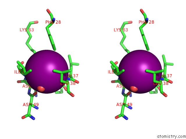
Iodine binding site 2 out of 12 in 3dhx
Go back to
Iodine binding site 2 out
of 12 in the Crystal Structure of Isolated C2 Domain of the Methionine Uptake Transporter
Mono view
Stereo pair view
Mono view
Stereo pair view
A full contact list of Iodine with other atoms in the I binding
site number 2 of Crystal Structure of Isolated C2 Domain of the Methionine Uptake Transporter within 5.0Å range:
|
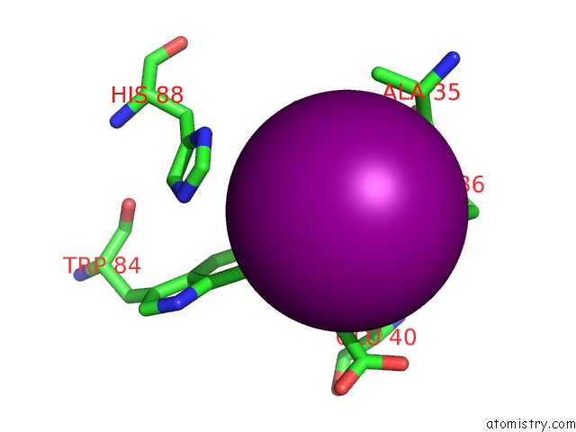
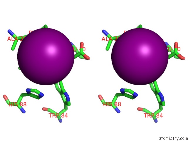
Iodine binding site 3 out of 12 in 3dhx
Go back to
Iodine binding site 3 out
of 12 in the Crystal Structure of Isolated C2 Domain of the Methionine Uptake Transporter
Mono view
Stereo pair view
Mono view
Stereo pair view
A full contact list of Iodine with other atoms in the I binding
site number 3 of Crystal Structure of Isolated C2 Domain of the Methionine Uptake Transporter within 5.0Å range:
|
Iodine binding site 4 out of 12 in 3dhx
Go back to
Iodine binding site 4 out
of 12 in the Crystal Structure of Isolated C2 Domain of the Methionine Uptake Transporter
Mono view
Stereo pair view
Mono view
Stereo pair view
A full contact list of Iodine with other atoms in the I binding
site number 4 of Crystal Structure of Isolated C2 Domain of the Methionine Uptake Transporter within 5.0Å range:
|
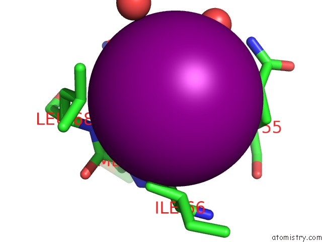
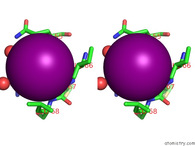
Iodine binding site 5 out of 12 in 3dhx
Go back to
Iodine binding site 5 out
of 12 in the Crystal Structure of Isolated C2 Domain of the Methionine Uptake Transporter
Mono view
Stereo pair view
Mono view
Stereo pair view
A full contact list of Iodine with other atoms in the I binding
site number 5 of Crystal Structure of Isolated C2 Domain of the Methionine Uptake Transporter within 5.0Å range:
|
Iodine binding site 6 out of 12 in 3dhx
Go back to
Iodine binding site 6 out
of 12 in the Crystal Structure of Isolated C2 Domain of the Methionine Uptake Transporter
Mono view
Stereo pair view
Mono view
Stereo pair view
A full contact list of Iodine with other atoms in the I binding
site number 6 of Crystal Structure of Isolated C2 Domain of the Methionine Uptake Transporter within 5.0Å range:
|
Iodine binding site 7 out of 12 in 3dhx
Go back to
Iodine binding site 7 out
of 12 in the Crystal Structure of Isolated C2 Domain of the Methionine Uptake Transporter
Mono view
Stereo pair view
Mono view
Stereo pair view
| A full contact list of Iodine with other atoms in the I binding site number 7 of Crystal Structure of Isolated C2 Domain of the Methionine Uptake Transporter within 5.0Å range: |
Iodine binding site 8 out of 12 in 3dhx
Go back to
Iodine binding site 8 out
of 12 in the Crystal Structure of Isolated C2 Domain of the Methionine Uptake Transporter
Mono view
Stereo pair view
Mono view
Stereo pair view
A full contact list of Iodine with other atoms in the I binding
site number 8 of Crystal Structure of Isolated C2 Domain of the Methionine Uptake Transporter within 5.0Å range:
|
Iodine binding site 9 out of 12 in 3dhx
Go back to
Iodine binding site 9 out
of 12 in the Crystal Structure of Isolated C2 Domain of the Methionine Uptake Transporter
Mono view
Stereo pair view
Mono view
Stereo pair view
A full contact list of Iodine with other atoms in the I binding
site number 9 of Crystal Structure of Isolated C2 Domain of the Methionine Uptake Transporter within 5.0Å range:
|
Iodine binding site 10 out of 12 in 3dhx
Go back to
Iodine binding site 10 out
of 12 in the Crystal Structure of Isolated C2 Domain of the Methionine Uptake Transporter
Mono view
Stereo pair view
Mono view
Stereo pair view
A full contact list of Iodine with other atoms in the I binding
site number 10 of Crystal Structure of Isolated C2 Domain of the Methionine Uptake Transporter within 5.0Å range:
|
Reference:
N.S.Kadaba,
J.T.Kaiser,
E.Johnson,
A.Lee,
D.C.Rees.
The High-Affinity E. Coli Methionine Abc Transporter: Structure and Allosteric Regulation. Science V. 321 250 2008.
ISSN: ISSN 0036-8075
PubMed: 18621668
DOI: 10.1126/SCIENCE.1157987
Page generated: Sun Aug 11 14:58:54 2024
ISSN: ISSN 0036-8075
PubMed: 18621668
DOI: 10.1126/SCIENCE.1157987
Last articles
Zn in 9MJ5Zn in 9HNW
Zn in 9G0L
Zn in 9FNE
Zn in 9DZN
Zn in 9E0I
Zn in 9D32
Zn in 9DAK
Zn in 8ZXC
Zn in 8ZUF