Iodine »
PDB 3cjq-3fnl »
3eqb »
Iodine in PDB 3eqb: X-Ray Structure of the Human Mitogen-Activated Protein Kinase Kinase 1 (MEK1) in A Complex with Ligand and Mgatp
Enzymatic activity of X-Ray Structure of the Human Mitogen-Activated Protein Kinase Kinase 1 (MEK1) in A Complex with Ligand and Mgatp
All present enzymatic activity of X-Ray Structure of the Human Mitogen-Activated Protein Kinase Kinase 1 (MEK1) in A Complex with Ligand and Mgatp:
2.7.12.2;
2.7.12.2;
Protein crystallography data
The structure of X-Ray Structure of the Human Mitogen-Activated Protein Kinase Kinase 1 (MEK1) in A Complex with Ligand and Mgatp, PDB code: 3eqb
was solved by
J.F.Ohren,
A.Pavlovsky,
E.Zhang,
with X-Ray Crystallography technique. A brief refinement statistics is given in the table below:
| Resolution Low / High (Å) | 20.00 / 2.62 |
| Space group | P 62 |
| Cell size a, b, c (Å), α, β, γ (°) | 82.128, 82.128, 129.099, 90.00, 90.00, 120.00 |
| R / Rfree (%) | 21 / 26.4 |
Other elements in 3eqb:
The structure of X-Ray Structure of the Human Mitogen-Activated Protein Kinase Kinase 1 (MEK1) in A Complex with Ligand and Mgatp also contains other interesting chemical elements:
| Fluorine | (F) | 3 atoms |
| Magnesium | (Mg) | 1 atom |
Iodine Binding Sites:
The binding sites of Iodine atom in the X-Ray Structure of the Human Mitogen-Activated Protein Kinase Kinase 1 (MEK1) in A Complex with Ligand and Mgatp
(pdb code 3eqb). This binding sites where shown within
5.0 Angstroms radius around Iodine atom.
In total only one binding site of Iodine was determined in the X-Ray Structure of the Human Mitogen-Activated Protein Kinase Kinase 1 (MEK1) in A Complex with Ligand and Mgatp, PDB code: 3eqb:
In total only one binding site of Iodine was determined in the X-Ray Structure of the Human Mitogen-Activated Protein Kinase Kinase 1 (MEK1) in A Complex with Ligand and Mgatp, PDB code: 3eqb:
Iodine binding site 1 out of 1 in 3eqb
Go back to
Iodine binding site 1 out
of 1 in the X-Ray Structure of the Human Mitogen-Activated Protein Kinase Kinase 1 (MEK1) in A Complex with Ligand and Mgatp
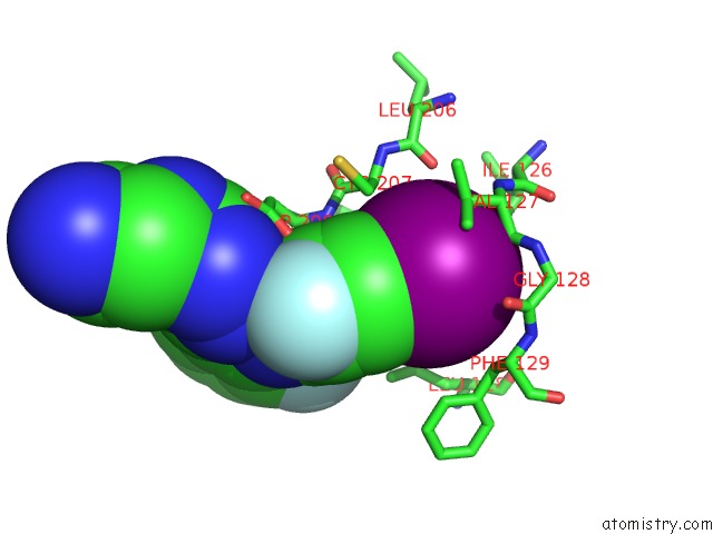
Mono view
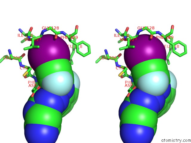
Stereo pair view
Mono view
Stereo pair view
A full contact list of Iodine with other atoms in the I binding
site number 1 of X-Ray Structure of the Human Mitogen-Activated Protein Kinase Kinase 1 (MEK1) in A Complex with Ligand and Mgatp within 5.0Å range:
|
Reference:
J.S.Warmus,
C.Flamme,
L.Y.Zhang,
S.Barrett,
A.Bridges,
H.Chen,
R.Gowan,
M.Kaufman,
J.Sebolt-Leopold,
W.Leopold,
R.Merriman,
J.Ohren,
A.Pavlovsky,
S.Przybranowski,
H.Tecle,
H.Valik,
C.Whitehead,
E.Zhang.
2-Alkylamino- and Alkoxy-Substituted 2-Amino-1,3,4-Oxadiazoles-O-Alkyl Benzohydroxamate Esters Replacements Retain the Desired Inhibition and Selectivity Against Mek (Map Erk Kinase). Bioorg.Med.Chem.Lett. V. 18 6171 2008.
ISSN: ISSN 0960-894X
PubMed: 18951019
DOI: 10.1016/J.BMCL.2008.10.015
Page generated: Sun Aug 11 15:06:14 2024
ISSN: ISSN 0960-894X
PubMed: 18951019
DOI: 10.1016/J.BMCL.2008.10.015
Last articles
Zn in 9JYWZn in 9IR4
Zn in 9IR3
Zn in 9GMX
Zn in 9GMW
Zn in 9JEJ
Zn in 9ERF
Zn in 9ERE
Zn in 9EGV
Zn in 9EGW