Iodine »
PDB 3cjq-3fnl »
3fgw »
Iodine in PDB 3fgw: One Chain Form of the 66.3 kDa Protein
Protein crystallography data
The structure of One Chain Form of the 66.3 kDa Protein, PDB code: 3fgw
was solved by
K.Lakomek,
A.Dickmanns,
R.Ficner,
with X-Ray Crystallography technique. A brief refinement statistics is given in the table below:
| Resolution Low / High (Å) | 46.07 / 2.80 |
| Space group | C 1 2 1 |
| Cell size a, b, c (Å), α, β, γ (°) | 146.690, 88.110, 73.550, 90.00, 111.10, 90.00 |
| R / Rfree (%) | 22.3 / 24.9 |
Other elements in 3fgw:
The structure of One Chain Form of the 66.3 kDa Protein also contains other interesting chemical elements:
| Sodium | (Na) | 1 atom |
Iodine Binding Sites:
The binding sites of Iodine atom in the One Chain Form of the 66.3 kDa Protein
(pdb code 3fgw). This binding sites where shown within
5.0 Angstroms radius around Iodine atom.
In total 7 binding sites of Iodine where determined in the One Chain Form of the 66.3 kDa Protein, PDB code: 3fgw:
Jump to Iodine binding site number: 1; 2; 3; 4; 5; 6; 7;
In total 7 binding sites of Iodine where determined in the One Chain Form of the 66.3 kDa Protein, PDB code: 3fgw:
Jump to Iodine binding site number: 1; 2; 3; 4; 5; 6; 7;
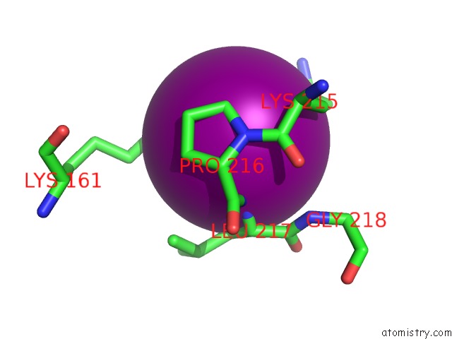
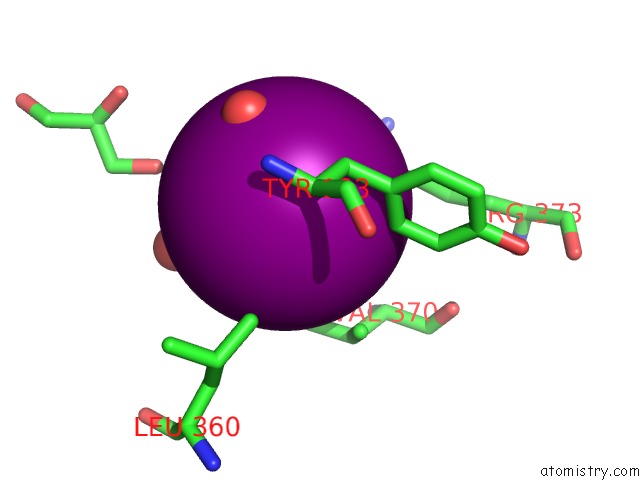
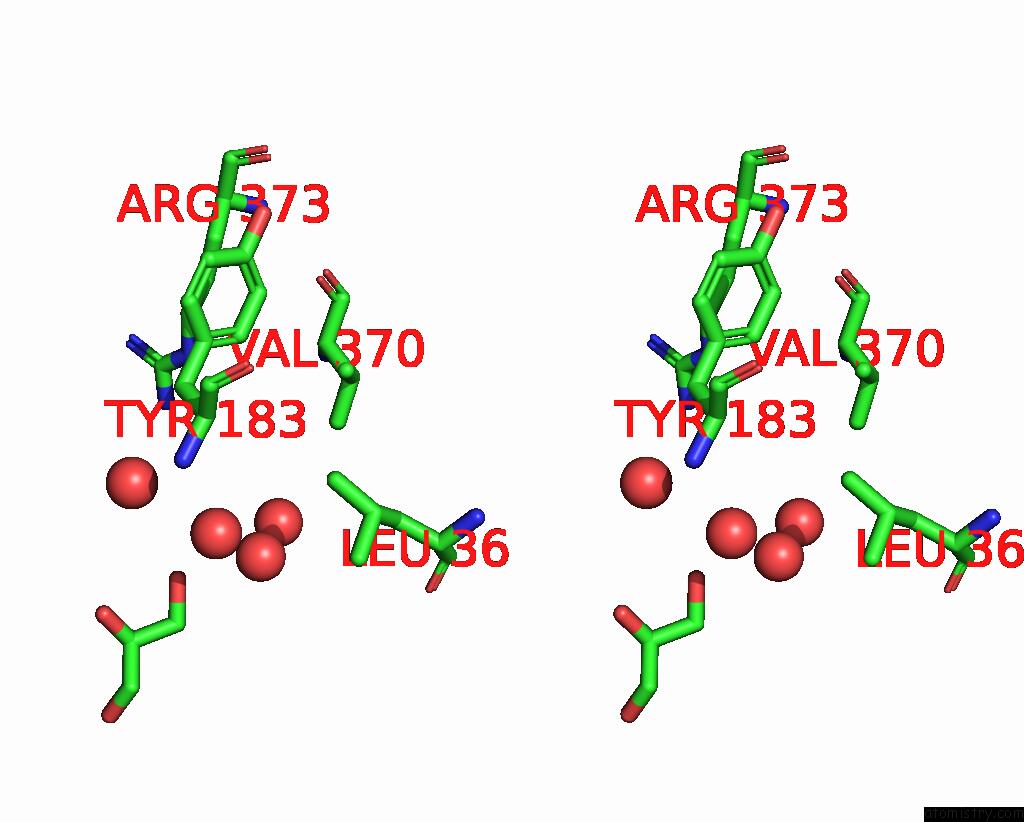
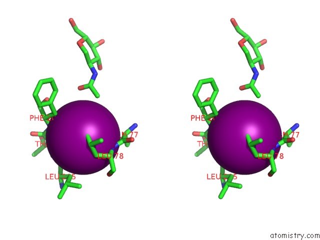
Iodine binding site 1 out of 7 in 3fgw
Go back to
Iodine binding site 1 out
of 7 in the One Chain Form of the 66.3 kDa Protein
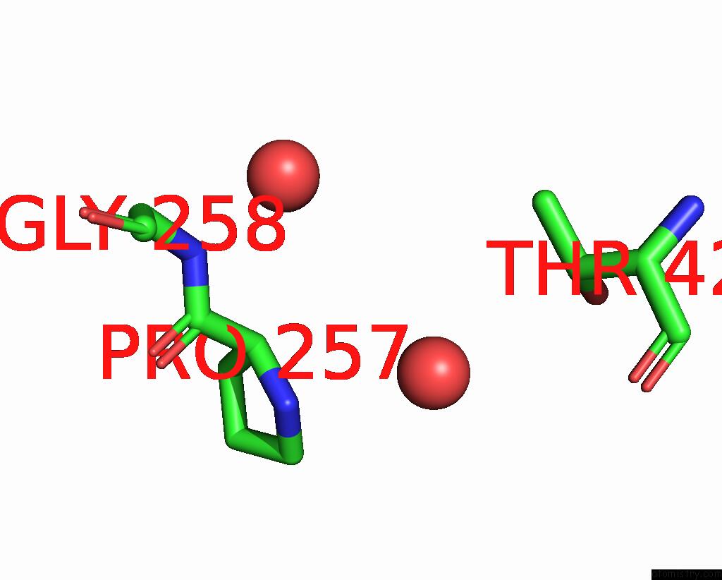
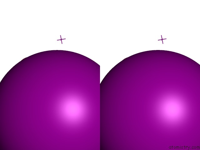
Mono view
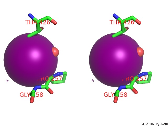
Stereo pair view
Mono view
Stereo pair view
A full contact list of Iodine with other atoms in the I binding
site number 1 of One Chain Form of the 66.3 kDa Protein within 5.0Å range:
|
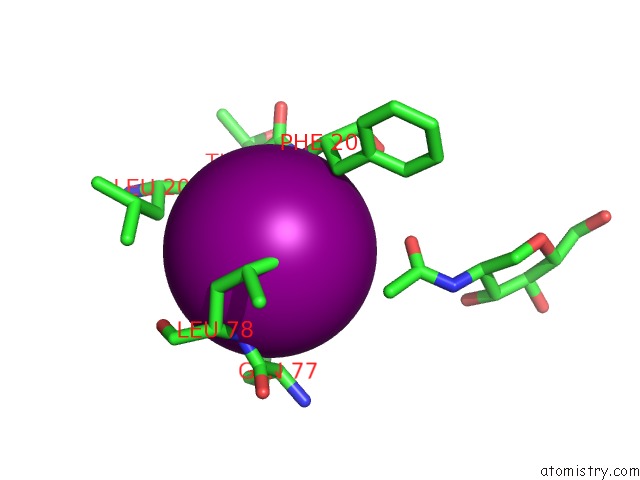
Iodine binding site 2 out of 7 in 3fgw
Go back to
Iodine binding site 2 out
of 7 in the One Chain Form of the 66.3 kDa Protein
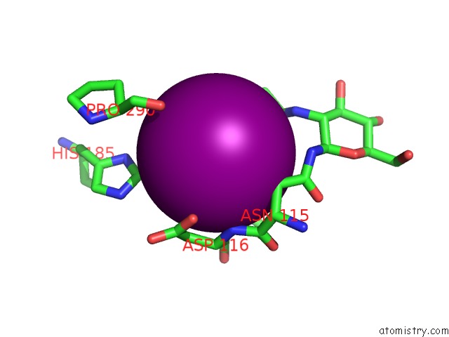
Mono view
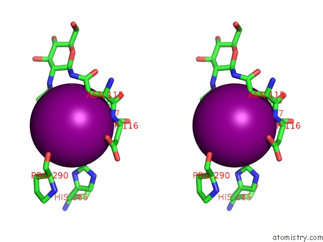
Stereo pair view
Mono view
Stereo pair view
A full contact list of Iodine with other atoms in the I binding
site number 2 of One Chain Form of the 66.3 kDa Protein within 5.0Å range:
|
Iodine binding site 3 out of 7 in 3fgw
Go back to
Iodine binding site 3 out
of 7 in the One Chain Form of the 66.3 kDa Protein
Mono view

Stereo pair view
Mono view
Stereo pair view
A full contact list of Iodine with other atoms in the I binding
site number 3 of One Chain Form of the 66.3 kDa Protein within 5.0Å range:
|
Iodine binding site 4 out of 7 in 3fgw
Go back to
Iodine binding site 4 out
of 7 in the One Chain Form of the 66.3 kDa Protein
Mono view
Stereo pair view
Mono view
Stereo pair view
A full contact list of Iodine with other atoms in the I binding
site number 4 of One Chain Form of the 66.3 kDa Protein within 5.0Å range:
|
Iodine binding site 5 out of 7 in 3fgw
Go back to
Iodine binding site 5 out
of 7 in the One Chain Form of the 66.3 kDa Protein
Mono view
Stereo pair view
Mono view
Stereo pair view
A full contact list of Iodine with other atoms in the I binding
site number 5 of One Chain Form of the 66.3 kDa Protein within 5.0Å range:
|
Iodine binding site 6 out of 7 in 3fgw
Go back to
Iodine binding site 6 out
of 7 in the One Chain Form of the 66.3 kDa Protein
Mono view
Stereo pair view
Mono view
Stereo pair view
A full contact list of Iodine with other atoms in the I binding
site number 6 of One Chain Form of the 66.3 kDa Protein within 5.0Å range:
|
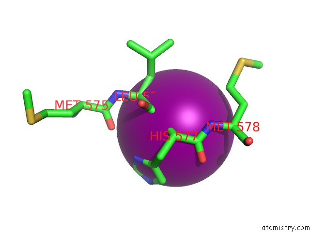
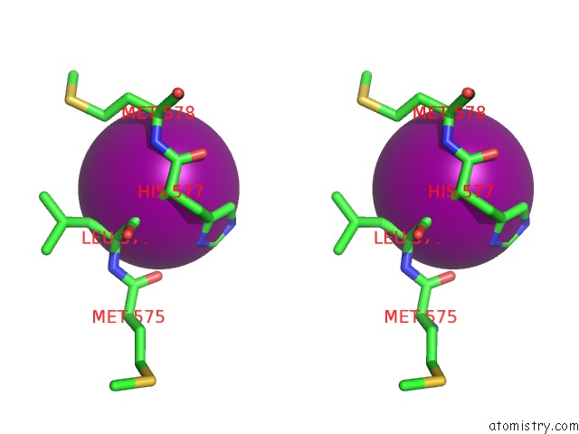
Iodine binding site 7 out of 7 in 3fgw
Go back to
Iodine binding site 7 out
of 7 in the One Chain Form of the 66.3 kDa Protein
Mono view
Stereo pair view
Mono view
Stereo pair view
A full contact list of Iodine with other atoms in the I binding
site number 7 of One Chain Form of the 66.3 kDa Protein within 5.0Å range:
|
Reference:
K.Lakomek,
A.Dickmanns,
M.Kettwig,
H.Urlaub,
R.Ficner,
T.Luebke.
Initial Insight Into the Function of the Lysosomal 66.3 kDa Protein From Mouse By Means of X-Ray Crystallography Bmc Struct.Biol. V. 9 56 2009.
ISSN: ESSN 1472-6807
PubMed: 19706171
DOI: 10.1186/1472-6807-9-56
Page generated: Fri Aug 8 14:15:29 2025
ISSN: ESSN 1472-6807
PubMed: 19706171
DOI: 10.1186/1472-6807-9-56
Last articles
I in 5KR8I in 5KM4
I in 5JYD
I in 5K2X
I in 5KIO
I in 5KIM
I in 5K6U
I in 5KC1
I in 5K7P
I in 5JVF