Iodine »
PDB 3g2t-3kxf »
3gcj »
Iodine in PDB 3gcj: Mode of Ligand Binding and Assignment of Subsites in Mammalian Peroxidases: Crystal Structure of Lactoperoxidase Complexes with Acetyl Salycylic Acid, Salicylhydroxamic Acid and Benzylhydroxamic Acid
Enzymatic activity of Mode of Ligand Binding and Assignment of Subsites in Mammalian Peroxidases: Crystal Structure of Lactoperoxidase Complexes with Acetyl Salycylic Acid, Salicylhydroxamic Acid and Benzylhydroxamic Acid
All present enzymatic activity of Mode of Ligand Binding and Assignment of Subsites in Mammalian Peroxidases: Crystal Structure of Lactoperoxidase Complexes with Acetyl Salycylic Acid, Salicylhydroxamic Acid and Benzylhydroxamic Acid:
1.11.1.7;
1.11.1.7;
Protein crystallography data
The structure of Mode of Ligand Binding and Assignment of Subsites in Mammalian Peroxidases: Crystal Structure of Lactoperoxidase Complexes with Acetyl Salycylic Acid, Salicylhydroxamic Acid and Benzylhydroxamic Acid, PDB code: 3gcj
was solved by
A.K.Singh,
N.Singh,
M.Sinha,
P.Kaur,
A.Srinivasan,
S.Sharma,
T.P.Singh,
with X-Ray Crystallography technique. A brief refinement statistics is given in the table below:
| Resolution Low / High (Å) | 19.47 / 2.34 |
| Space group | P 1 21 1 |
| Cell size a, b, c (Å), α, β, γ (°) | 54.618, 80.553, 77.803, 90.00, 102.55, 90.00 |
| R / Rfree (%) | 20.4 / 21.2 |
Other elements in 3gcj:
The structure of Mode of Ligand Binding and Assignment of Subsites in Mammalian Peroxidases: Crystal Structure of Lactoperoxidase Complexes with Acetyl Salycylic Acid, Salicylhydroxamic Acid and Benzylhydroxamic Acid also contains other interesting chemical elements:
| Iron | (Fe) | 1 atom |
| Calcium | (Ca) | 1 atom |
Iodine Binding Sites:
The binding sites of Iodine atom in the Mode of Ligand Binding and Assignment of Subsites in Mammalian Peroxidases: Crystal Structure of Lactoperoxidase Complexes with Acetyl Salycylic Acid, Salicylhydroxamic Acid and Benzylhydroxamic Acid
(pdb code 3gcj). This binding sites where shown within
5.0 Angstroms radius around Iodine atom.
In total 8 binding sites of Iodine where determined in the Mode of Ligand Binding and Assignment of Subsites in Mammalian Peroxidases: Crystal Structure of Lactoperoxidase Complexes with Acetyl Salycylic Acid, Salicylhydroxamic Acid and Benzylhydroxamic Acid, PDB code: 3gcj:
Jump to Iodine binding site number: 1; 2; 3; 4; 5; 6; 7; 8;
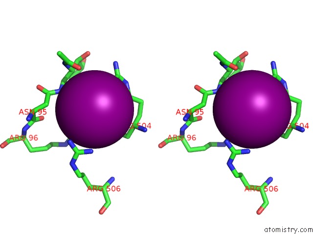
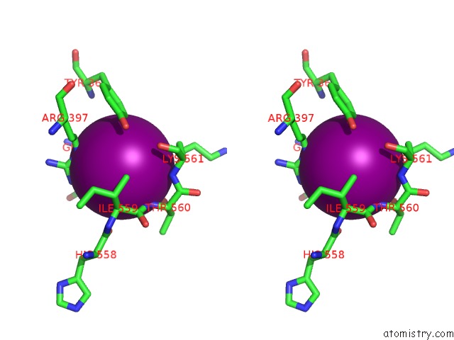
Iodine binding site 1 out of 8 in 3gcj
Go back to
Iodine binding site 1 out
of 8 in the Mode of Ligand Binding and Assignment of Subsites in Mammalian Peroxidases: Crystal Structure of Lactoperoxidase Complexes with Acetyl Salycylic Acid, Salicylhydroxamic Acid and Benzylhydroxamic Acid

Mono view
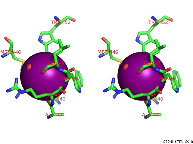
Stereo pair view
Mono view
Stereo pair view
A full contact list of Iodine with other atoms in the I binding
site number 1 of Mode of Ligand Binding and Assignment of Subsites in Mammalian Peroxidases: Crystal Structure of Lactoperoxidase Complexes with Acetyl Salycylic Acid, Salicylhydroxamic Acid and Benzylhydroxamic Acid within 5.0Å range:
|
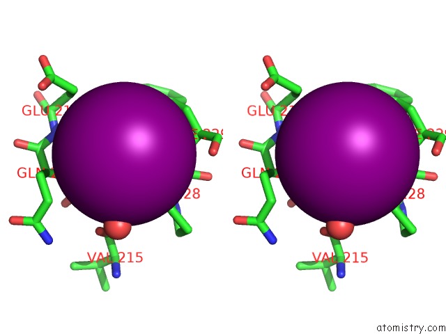
Iodine binding site 2 out of 8 in 3gcj
Go back to
Iodine binding site 2 out
of 8 in the Mode of Ligand Binding and Assignment of Subsites in Mammalian Peroxidases: Crystal Structure of Lactoperoxidase Complexes with Acetyl Salycylic Acid, Salicylhydroxamic Acid and Benzylhydroxamic Acid
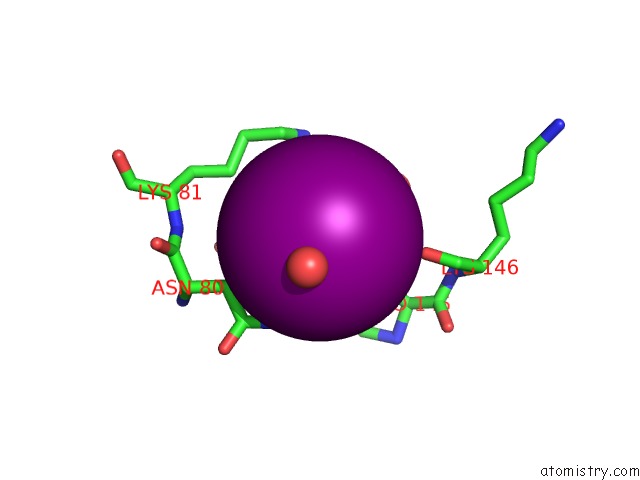
Mono view
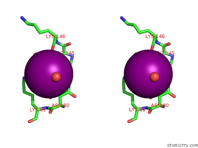
Stereo pair view
Mono view
Stereo pair view
A full contact list of Iodine with other atoms in the I binding
site number 2 of Mode of Ligand Binding and Assignment of Subsites in Mammalian Peroxidases: Crystal Structure of Lactoperoxidase Complexes with Acetyl Salycylic Acid, Salicylhydroxamic Acid and Benzylhydroxamic Acid within 5.0Å range:
|
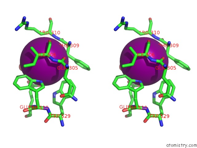
Iodine binding site 3 out of 8 in 3gcj
Go back to
Iodine binding site 3 out
of 8 in the Mode of Ligand Binding and Assignment of Subsites in Mammalian Peroxidases: Crystal Structure of Lactoperoxidase Complexes with Acetyl Salycylic Acid, Salicylhydroxamic Acid and Benzylhydroxamic Acid
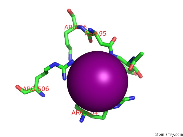
Mono view
Stereo pair view
Mono view
Stereo pair view
A full contact list of Iodine with other atoms in the I binding
site number 3 of Mode of Ligand Binding and Assignment of Subsites in Mammalian Peroxidases: Crystal Structure of Lactoperoxidase Complexes with Acetyl Salycylic Acid, Salicylhydroxamic Acid and Benzylhydroxamic Acid within 5.0Å range:
|
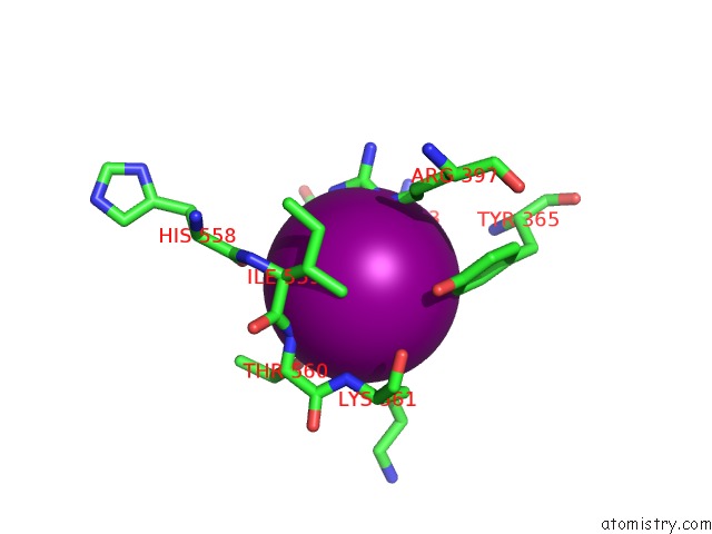
Iodine binding site 4 out of 8 in 3gcj
Go back to
Iodine binding site 4 out
of 8 in the Mode of Ligand Binding and Assignment of Subsites in Mammalian Peroxidases: Crystal Structure of Lactoperoxidase Complexes with Acetyl Salycylic Acid, Salicylhydroxamic Acid and Benzylhydroxamic Acid

Mono view
Stereo pair view
Mono view
Stereo pair view
A full contact list of Iodine with other atoms in the I binding
site number 4 of Mode of Ligand Binding and Assignment of Subsites in Mammalian Peroxidases: Crystal Structure of Lactoperoxidase Complexes with Acetyl Salycylic Acid, Salicylhydroxamic Acid and Benzylhydroxamic Acid within 5.0Å range:
|
Iodine binding site 5 out of 8 in 3gcj
Go back to
Iodine binding site 5 out
of 8 in the Mode of Ligand Binding and Assignment of Subsites in Mammalian Peroxidases: Crystal Structure of Lactoperoxidase Complexes with Acetyl Salycylic Acid, Salicylhydroxamic Acid and Benzylhydroxamic Acid

Mono view
Stereo pair view
Mono view
Stereo pair view
A full contact list of Iodine with other atoms in the I binding
site number 5 of Mode of Ligand Binding and Assignment of Subsites in Mammalian Peroxidases: Crystal Structure of Lactoperoxidase Complexes with Acetyl Salycylic Acid, Salicylhydroxamic Acid and Benzylhydroxamic Acid within 5.0Å range:
|
Iodine binding site 6 out of 8 in 3gcj
Go back to
Iodine binding site 6 out
of 8 in the Mode of Ligand Binding and Assignment of Subsites in Mammalian Peroxidases: Crystal Structure of Lactoperoxidase Complexes with Acetyl Salycylic Acid, Salicylhydroxamic Acid and Benzylhydroxamic Acid
Mono view
Stereo pair view
Mono view
Stereo pair view
A full contact list of Iodine with other atoms in the I binding
site number 6 of Mode of Ligand Binding and Assignment of Subsites in Mammalian Peroxidases: Crystal Structure of Lactoperoxidase Complexes with Acetyl Salycylic Acid, Salicylhydroxamic Acid and Benzylhydroxamic Acid within 5.0Å range:
|
Iodine binding site 7 out of 8 in 3gcj
Go back to
Iodine binding site 7 out
of 8 in the Mode of Ligand Binding and Assignment of Subsites in Mammalian Peroxidases: Crystal Structure of Lactoperoxidase Complexes with Acetyl Salycylic Acid, Salicylhydroxamic Acid and Benzylhydroxamic Acid

Mono view
Stereo pair view
Mono view
Stereo pair view
A full contact list of Iodine with other atoms in the I binding
site number 7 of Mode of Ligand Binding and Assignment of Subsites in Mammalian Peroxidases: Crystal Structure of Lactoperoxidase Complexes with Acetyl Salycylic Acid, Salicylhydroxamic Acid and Benzylhydroxamic Acid within 5.0Å range:
|
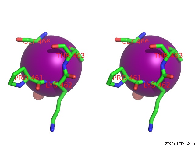
Iodine binding site 8 out of 8 in 3gcj
Go back to
Iodine binding site 8 out
of 8 in the Mode of Ligand Binding and Assignment of Subsites in Mammalian Peroxidases: Crystal Structure of Lactoperoxidase Complexes with Acetyl Salycylic Acid, Salicylhydroxamic Acid and Benzylhydroxamic Acid

Mono view

Stereo pair view
Mono view
Stereo pair view
A full contact list of Iodine with other atoms in the I binding
site number 8 of Mode of Ligand Binding and Assignment of Subsites in Mammalian Peroxidases: Crystal Structure of Lactoperoxidase Complexes with Acetyl Salycylic Acid, Salicylhydroxamic Acid and Benzylhydroxamic Acid within 5.0Å range:
|
Reference:
A.K.Singh,
N.Singh,
M.Sinha,
P.Kaur,
A.Srinivasan,
S.Sharma,
T.P.Singh.
Mode of Ligand Binding and Assignment of Subsites in Mammalian Peroxidases: Crystal Structure of Lactoperoxidase Complexes with Acetyl Salycylic Acid, Salicylhydroxamic Acid and Benzylhydroxamic Acid To Be Published.
Page generated: Fri Aug 8 14:17:39 2025
Last articles
I in 5M1YI in 5MPH
I in 5MJO
I in 5MHP
I in 5MA2
I in 5MHG
I in 5MHE
I in 5MBG
I in 5M0S
I in 5M0M