Iodine »
PDB 3g2t-3kxf »
3gh8 »
Iodine in PDB 3gh8: Crystal Structure of Mus Musculus Iodotyrosine Deiodinase (Iyd) Bound to Fmn and Di-Iodotyrosine (Dit)
Protein crystallography data
The structure of Crystal Structure of Mus Musculus Iodotyrosine Deiodinase (Iyd) Bound to Fmn and Di-Iodotyrosine (Dit), PDB code: 3gh8
was solved by
S.R.Thomas,
P.M.Mctamney,
J.M.Adler,
N.Laronde-Leblanc,
S.E.Rokita,
with X-Ray Crystallography technique. A brief refinement statistics is given in the table below:
| Resolution Low / High (Å) | 30.00 / 2.61 |
| Space group | P 1 21 1 |
| Cell size a, b, c (Å), α, β, γ (°) | 50.610, 112.567, 189.253, 90.00, 89.92, 90.00 |
| R / Rfree (%) | 18.1 / 26.4 |
Iodine Binding Sites:
Pages:
>>> Page 1 <<< Page 2, Binding sites: 11 - 16;Binding sites:
The binding sites of Iodine atom in the Crystal Structure of Mus Musculus Iodotyrosine Deiodinase (Iyd) Bound to Fmn and Di-Iodotyrosine (Dit) (pdb code 3gh8). This binding sites where shown within 5.0 Angstroms radius around Iodine atom.In total 16 binding sites of Iodine where determined in the Crystal Structure of Mus Musculus Iodotyrosine Deiodinase (Iyd) Bound to Fmn and Di-Iodotyrosine (Dit), PDB code: 3gh8:
Jump to Iodine binding site number: 1; 2; 3; 4; 5; 6; 7; 8; 9; 10;
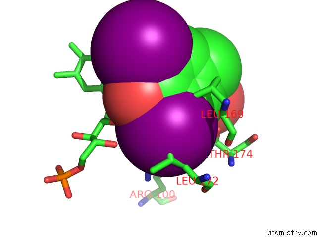
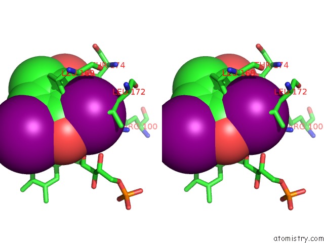
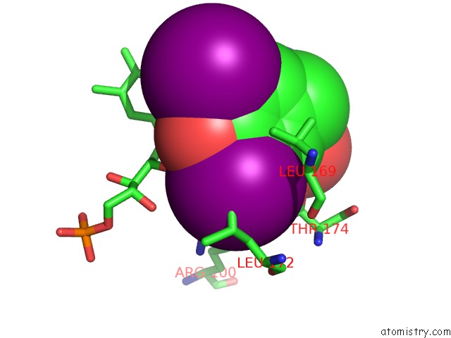
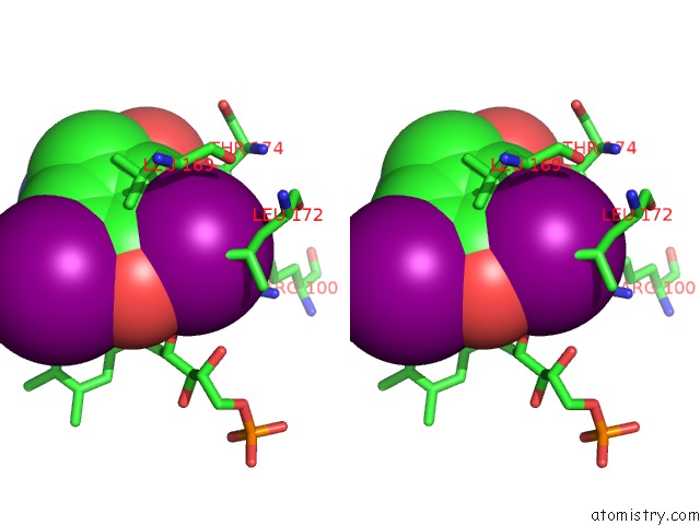
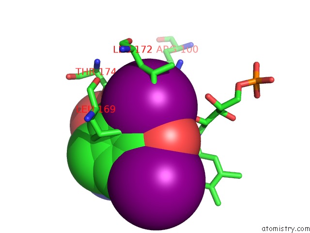
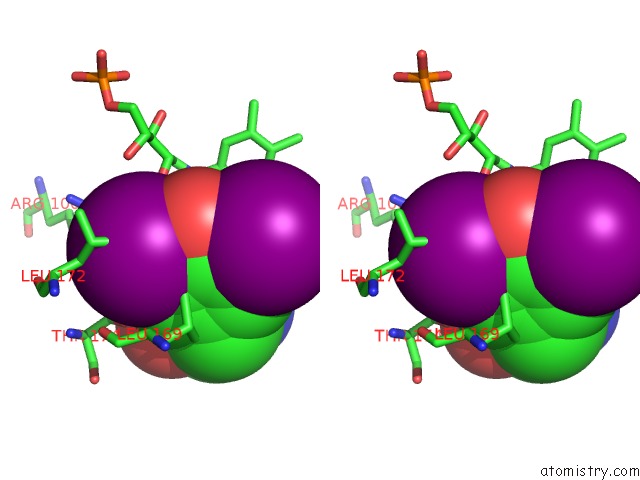
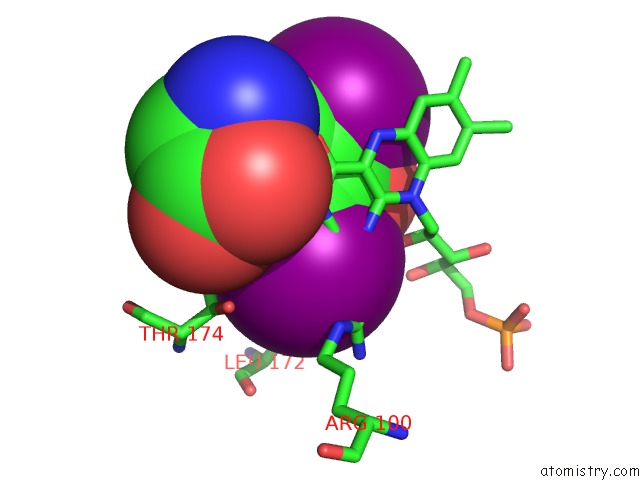
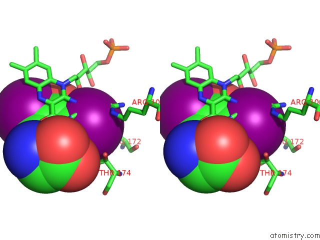
Iodine binding site 1 out of 16 in 3gh8
Go back to
Iodine binding site 1 out
of 16 in the Crystal Structure of Mus Musculus Iodotyrosine Deiodinase (Iyd) Bound to Fmn and Di-Iodotyrosine (Dit)
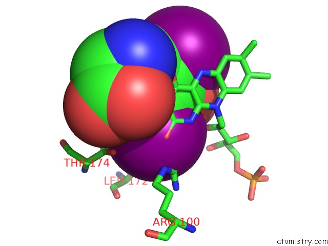
Mono view
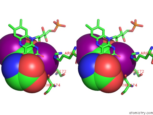
Stereo pair view
Mono view
Stereo pair view
A full contact list of Iodine with other atoms in the I binding
site number 1 of Crystal Structure of Mus Musculus Iodotyrosine Deiodinase (Iyd) Bound to Fmn and Di-Iodotyrosine (Dit) within 5.0Å range:
|
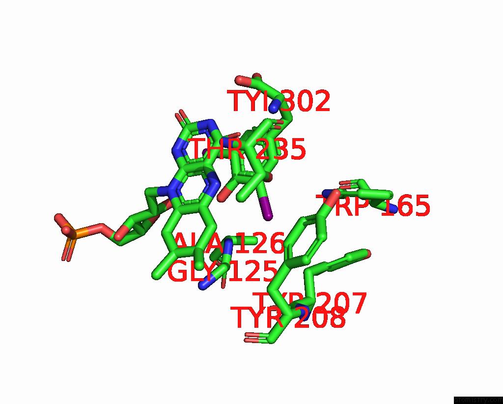
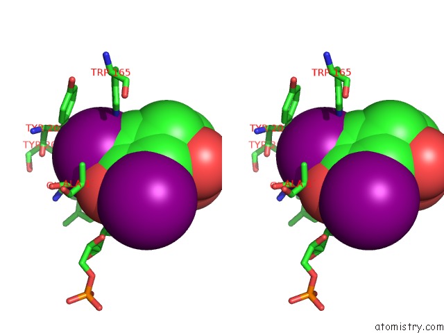
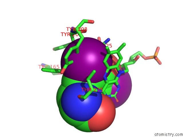
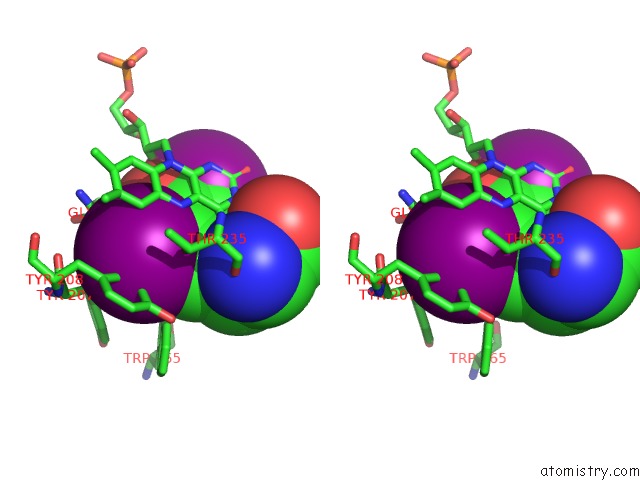
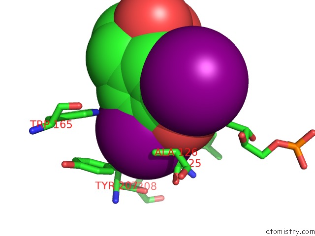
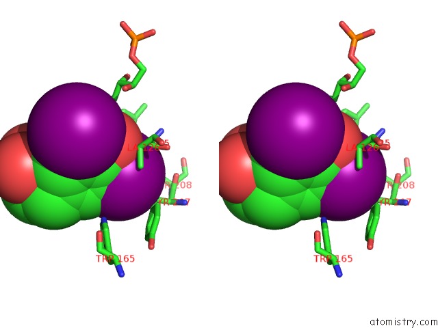
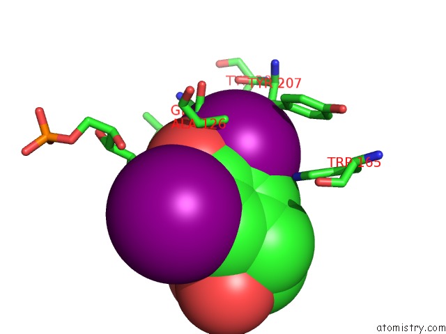
Iodine binding site 2 out of 16 in 3gh8
Go back to
Iodine binding site 2 out
of 16 in the Crystal Structure of Mus Musculus Iodotyrosine Deiodinase (Iyd) Bound to Fmn and Di-Iodotyrosine (Dit)
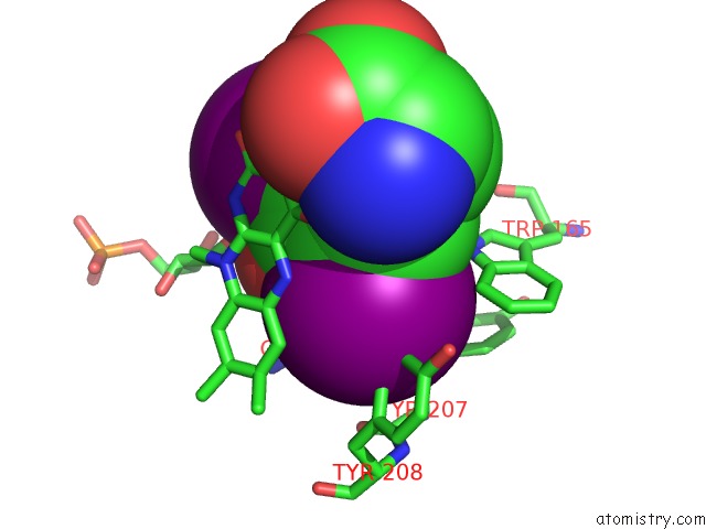
Mono view
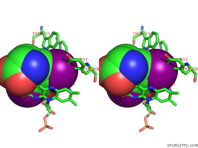
Stereo pair view
Mono view
Stereo pair view
A full contact list of Iodine with other atoms in the I binding
site number 2 of Crystal Structure of Mus Musculus Iodotyrosine Deiodinase (Iyd) Bound to Fmn and Di-Iodotyrosine (Dit) within 5.0Å range:
|
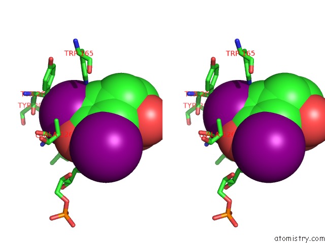
Iodine binding site 3 out of 16 in 3gh8
Go back to
Iodine binding site 3 out
of 16 in the Crystal Structure of Mus Musculus Iodotyrosine Deiodinase (Iyd) Bound to Fmn and Di-Iodotyrosine (Dit)
Mono view
Stereo pair view
Mono view
Stereo pair view
A full contact list of Iodine with other atoms in the I binding
site number 3 of Crystal Structure of Mus Musculus Iodotyrosine Deiodinase (Iyd) Bound to Fmn and Di-Iodotyrosine (Dit) within 5.0Å range:
|
Iodine binding site 4 out of 16 in 3gh8
Go back to
Iodine binding site 4 out
of 16 in the Crystal Structure of Mus Musculus Iodotyrosine Deiodinase (Iyd) Bound to Fmn and Di-Iodotyrosine (Dit)
Mono view
Stereo pair view
Mono view
Stereo pair view
A full contact list of Iodine with other atoms in the I binding
site number 4 of Crystal Structure of Mus Musculus Iodotyrosine Deiodinase (Iyd) Bound to Fmn and Di-Iodotyrosine (Dit) within 5.0Å range:
|
Iodine binding site 5 out of 16 in 3gh8
Go back to
Iodine binding site 5 out
of 16 in the Crystal Structure of Mus Musculus Iodotyrosine Deiodinase (Iyd) Bound to Fmn and Di-Iodotyrosine (Dit)
Mono view
Stereo pair view
Mono view
Stereo pair view
A full contact list of Iodine with other atoms in the I binding
site number 5 of Crystal Structure of Mus Musculus Iodotyrosine Deiodinase (Iyd) Bound to Fmn and Di-Iodotyrosine (Dit) within 5.0Å range:
|
Iodine binding site 6 out of 16 in 3gh8
Go back to
Iodine binding site 6 out
of 16 in the Crystal Structure of Mus Musculus Iodotyrosine Deiodinase (Iyd) Bound to Fmn and Di-Iodotyrosine (Dit)
Mono view
Stereo pair view
Mono view
Stereo pair view
A full contact list of Iodine with other atoms in the I binding
site number 6 of Crystal Structure of Mus Musculus Iodotyrosine Deiodinase (Iyd) Bound to Fmn and Di-Iodotyrosine (Dit) within 5.0Å range:
|
Iodine binding site 7 out of 16 in 3gh8
Go back to
Iodine binding site 7 out
of 16 in the Crystal Structure of Mus Musculus Iodotyrosine Deiodinase (Iyd) Bound to Fmn and Di-Iodotyrosine (Dit)
Mono view
Stereo pair view
Mono view
Stereo pair view
A full contact list of Iodine with other atoms in the I binding
site number 7 of Crystal Structure of Mus Musculus Iodotyrosine Deiodinase (Iyd) Bound to Fmn and Di-Iodotyrosine (Dit) within 5.0Å range:
|
Iodine binding site 8 out of 16 in 3gh8
Go back to
Iodine binding site 8 out
of 16 in the Crystal Structure of Mus Musculus Iodotyrosine Deiodinase (Iyd) Bound to Fmn and Di-Iodotyrosine (Dit)
Mono view
Stereo pair view
Mono view
Stereo pair view
A full contact list of Iodine with other atoms in the I binding
site number 8 of Crystal Structure of Mus Musculus Iodotyrosine Deiodinase (Iyd) Bound to Fmn and Di-Iodotyrosine (Dit) within 5.0Å range:
|
Iodine binding site 9 out of 16 in 3gh8
Go back to
Iodine binding site 9 out
of 16 in the Crystal Structure of Mus Musculus Iodotyrosine Deiodinase (Iyd) Bound to Fmn and Di-Iodotyrosine (Dit)
Mono view
Stereo pair view
Mono view
Stereo pair view
A full contact list of Iodine with other atoms in the I binding
site number 9 of Crystal Structure of Mus Musculus Iodotyrosine Deiodinase (Iyd) Bound to Fmn and Di-Iodotyrosine (Dit) within 5.0Å range:
|
Iodine binding site 10 out of 16 in 3gh8
Go back to
Iodine binding site 10 out
of 16 in the Crystal Structure of Mus Musculus Iodotyrosine Deiodinase (Iyd) Bound to Fmn and Di-Iodotyrosine (Dit)
Mono view
Stereo pair view
Mono view
Stereo pair view
A full contact list of Iodine with other atoms in the I binding
site number 10 of Crystal Structure of Mus Musculus Iodotyrosine Deiodinase (Iyd) Bound to Fmn and Di-Iodotyrosine (Dit) within 5.0Å range:
|
Reference:
S.R.Thomas,
P.M.Mctamney,
J.M.Adler,
N.Laronde-Leblanc,
S.E.Rokita.
Crystal Structure of Iodotyrosine Deiodinase, A Novel Flavoprotein Responsible For Iodide Salvage in Thyroid Glands. J.Biol.Chem. V. 284 19659 2009.
ISSN: ISSN 0021-9258
PubMed: 19436071
DOI: 10.1074/JBC.M109.013458
Page generated: Sun Aug 11 15:16:26 2024
ISSN: ISSN 0021-9258
PubMed: 19436071
DOI: 10.1074/JBC.M109.013458
Last articles
Zn in 9J0NZn in 9J0O
Zn in 9J0P
Zn in 9FJX
Zn in 9EKB
Zn in 9C0F
Zn in 9CAH
Zn in 9CH0
Zn in 9CH3
Zn in 9CH1