Iodine »
PDB 3g2t-3kxf »
3gqx »
Iodine in PDB 3gqx: Pyrococcus Horikoshii NOP5 Rna Binding Domain From A Twinned Crystal Form
Protein crystallography data
The structure of Pyrococcus Horikoshii NOP5 Rna Binding Domain From A Twinned Crystal Form, PDB code: 3gqx
was solved by
F.E.Reyes,
J.W.Hardin,
R.T.Batey,
with X-Ray Crystallography technique. A brief refinement statistics is given in the table below:
| Resolution Low / High (Å) | 23.68 / 2.50 |
| Space group | P 41 |
| Cell size a, b, c (Å), α, β, γ (°) | 73.604, 73.604, 114.279, 90.00, 90.00, 90.00 |
| R / Rfree (%) | 25.2 / 28.7 |
Iodine Binding Sites:
The binding sites of Iodine atom in the Pyrococcus Horikoshii NOP5 Rna Binding Domain From A Twinned Crystal Form
(pdb code 3gqx). This binding sites where shown within
5.0 Angstroms radius around Iodine atom.
In total 3 binding sites of Iodine where determined in the Pyrococcus Horikoshii NOP5 Rna Binding Domain From A Twinned Crystal Form, PDB code: 3gqx:
Jump to Iodine binding site number: 1; 2; 3;
In total 3 binding sites of Iodine where determined in the Pyrococcus Horikoshii NOP5 Rna Binding Domain From A Twinned Crystal Form, PDB code: 3gqx:
Jump to Iodine binding site number: 1; 2; 3;
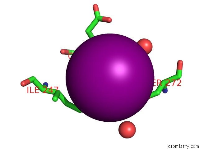
Iodine binding site 1 out of 3 in 3gqx
Go back to
Iodine binding site 1 out
of 3 in the Pyrococcus Horikoshii NOP5 Rna Binding Domain From A Twinned Crystal Form
Mono view

Stereo pair view
Mono view
Stereo pair view
A full contact list of Iodine with other atoms in the I binding
site number 1 of Pyrococcus Horikoshii NOP5 Rna Binding Domain From A Twinned Crystal Form within 5.0Å range:
|
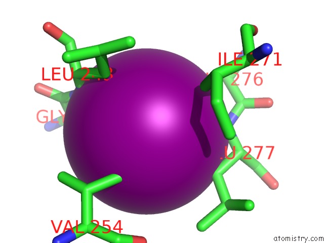
Iodine binding site 2 out of 3 in 3gqx
Go back to
Iodine binding site 2 out
of 3 in the Pyrococcus Horikoshii NOP5 Rna Binding Domain From A Twinned Crystal Form
Mono view

Stereo pair view
Mono view
Stereo pair view
A full contact list of Iodine with other atoms in the I binding
site number 2 of Pyrococcus Horikoshii NOP5 Rna Binding Domain From A Twinned Crystal Form within 5.0Å range:
|
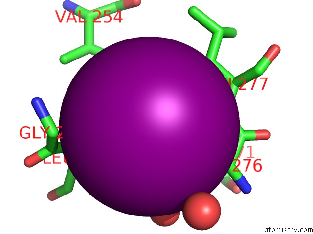
Iodine binding site 3 out of 3 in 3gqx
Go back to
Iodine binding site 3 out
of 3 in the Pyrococcus Horikoshii NOP5 Rna Binding Domain From A Twinned Crystal Form
Mono view
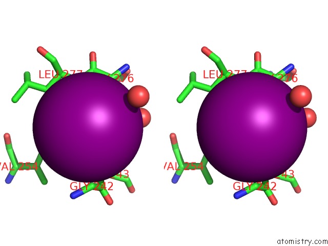
Stereo pair view
Mono view
Stereo pair view
A full contact list of Iodine with other atoms in the I binding
site number 3 of Pyrococcus Horikoshii NOP5 Rna Binding Domain From A Twinned Crystal Form within 5.0Å range:
|
Reference:
J.W.Hardin,
F.E.Reyes,
R.T.Batey.
Analysis of A Critical Interaction Within the Archaeal Box C/D Small Ribonucleoprotein Complex J.Biol.Chem. V. 284 15317 2009.
ISSN: ISSN 0021-9258
PubMed: 19336398
DOI: 10.1074/JBC.M901368200
Page generated: Sun Aug 11 15:16:42 2024
ISSN: ISSN 0021-9258
PubMed: 19336398
DOI: 10.1074/JBC.M901368200
Last articles
Zn in 9J0NZn in 9J0O
Zn in 9J0P
Zn in 9FJX
Zn in 9EKB
Zn in 9C0F
Zn in 9CAH
Zn in 9CH0
Zn in 9CH3
Zn in 9CH1