Iodine »
PDB 3g2t-3kxf »
3gt4 »
Iodine in PDB 3gt4: Structure of Proteinase K with the Magic Triangle I3C
Enzymatic activity of Structure of Proteinase K with the Magic Triangle I3C
All present enzymatic activity of Structure of Proteinase K with the Magic Triangle I3C:
3.4.21.64;
3.4.21.64;
Protein crystallography data
The structure of Structure of Proteinase K with the Magic Triangle I3C, PDB code: 3gt4
was solved by
T.Beck,
T.Gruene,
G.M.Sheldrick,
with X-Ray Crystallography technique. A brief refinement statistics is given in the table below:
| Resolution Low / High (Å) | 47.90 / 1.76 |
| Space group | P 43 21 2 |
| Cell size a, b, c (Å), α, β, γ (°) | 67.784, 67.784, 101.845, 90.00, 90.00, 90.00 |
| R / Rfree (%) | 14 / 19.1 |
Iodine Binding Sites:
The binding sites of Iodine atom in the Structure of Proteinase K with the Magic Triangle I3C
(pdb code 3gt4). This binding sites where shown within
5.0 Angstroms radius around Iodine atom.
In total 9 binding sites of Iodine where determined in the Structure of Proteinase K with the Magic Triangle I3C, PDB code: 3gt4:
Jump to Iodine binding site number: 1; 2; 3; 4; 5; 6; 7; 8; 9;
In total 9 binding sites of Iodine where determined in the Structure of Proteinase K with the Magic Triangle I3C, PDB code: 3gt4:
Jump to Iodine binding site number: 1; 2; 3; 4; 5; 6; 7; 8; 9;
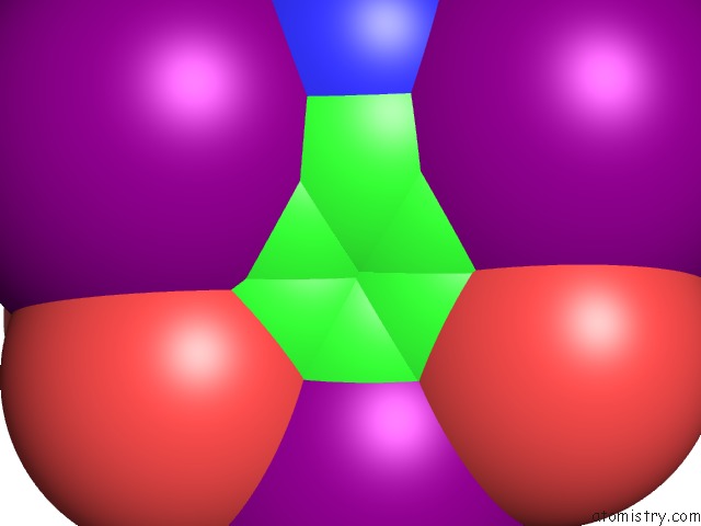
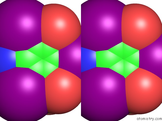
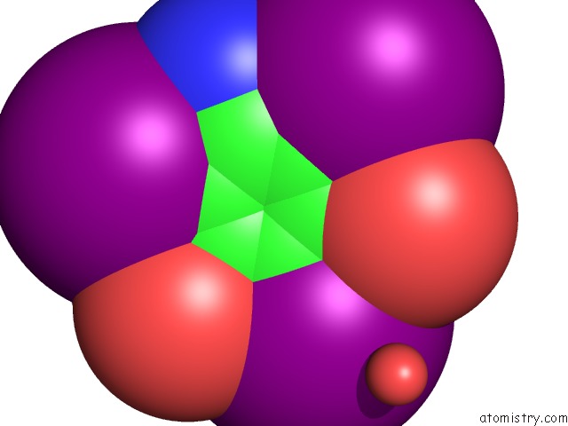
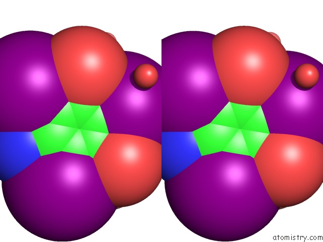
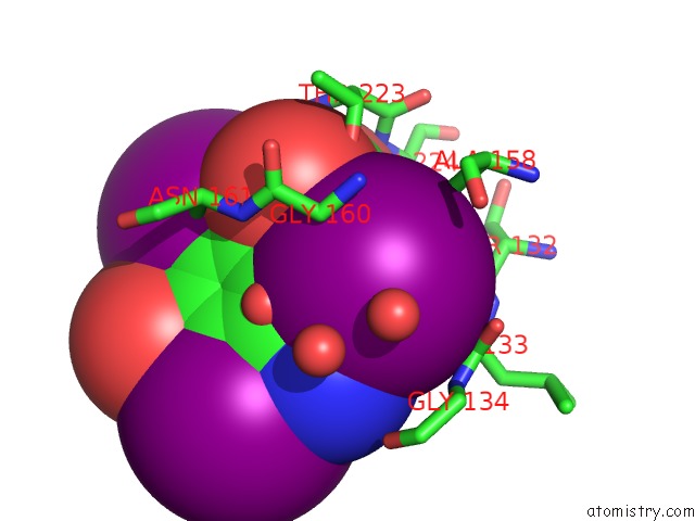
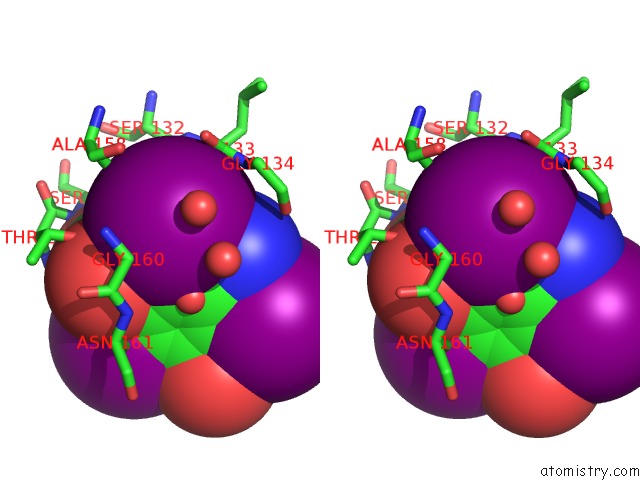
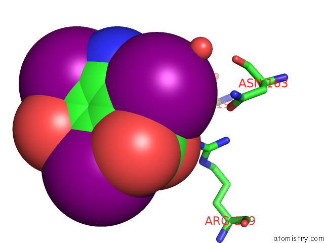
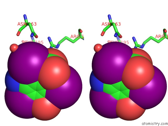
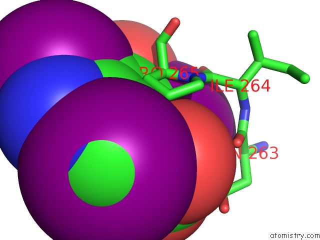
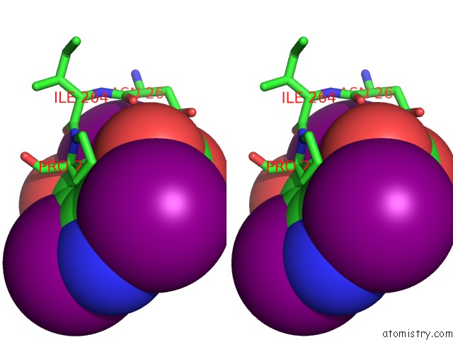
Iodine binding site 1 out of 9 in 3gt4
Go back to
Iodine binding site 1 out
of 9 in the Structure of Proteinase K with the Magic Triangle I3C
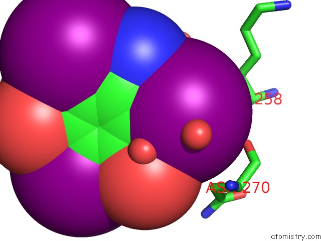
Mono view
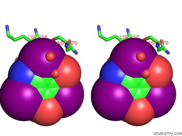
Stereo pair view
Mono view
Stereo pair view
A full contact list of Iodine with other atoms in the I binding
site number 1 of Structure of Proteinase K with the Magic Triangle I3C within 5.0Å range:
|
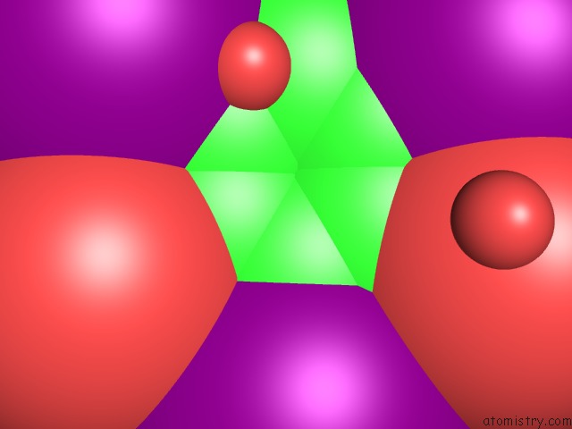
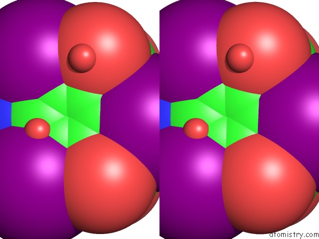
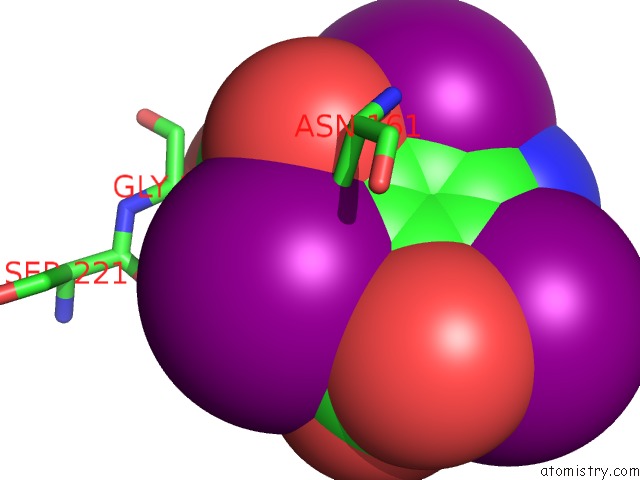
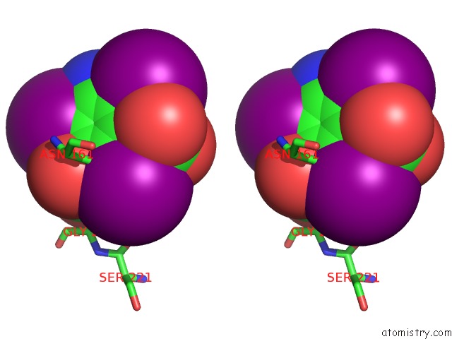
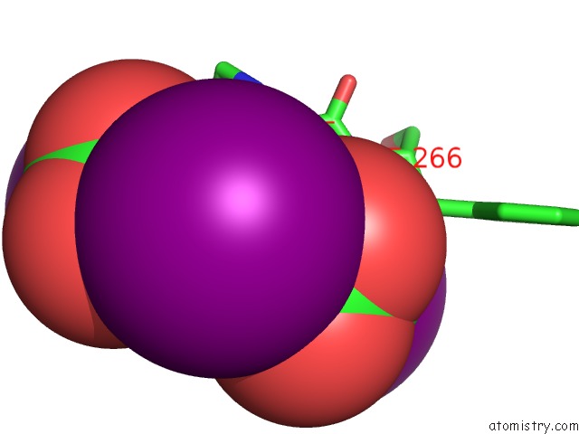
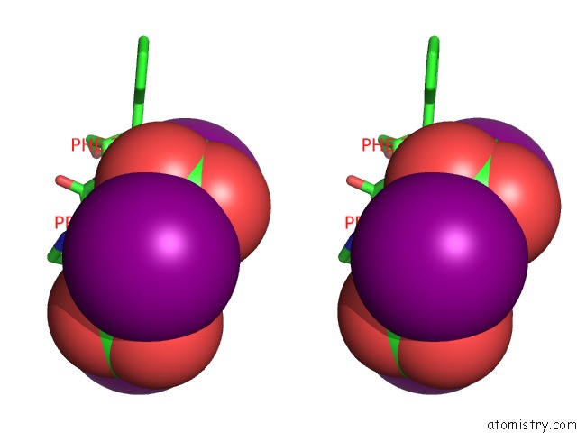
Iodine binding site 2 out of 9 in 3gt4
Go back to
Iodine binding site 2 out
of 9 in the Structure of Proteinase K with the Magic Triangle I3C
Mono view
Stereo pair view
Mono view
Stereo pair view
A full contact list of Iodine with other atoms in the I binding
site number 2 of Structure of Proteinase K with the Magic Triangle I3C within 5.0Å range:
|
Iodine binding site 3 out of 9 in 3gt4
Go back to
Iodine binding site 3 out
of 9 in the Structure of Proteinase K with the Magic Triangle I3C
Mono view
Stereo pair view
Mono view
Stereo pair view
A full contact list of Iodine with other atoms in the I binding
site number 3 of Structure of Proteinase K with the Magic Triangle I3C within 5.0Å range:
|
Iodine binding site 4 out of 9 in 3gt4
Go back to
Iodine binding site 4 out
of 9 in the Structure of Proteinase K with the Magic Triangle I3C
Mono view
Stereo pair view
Mono view
Stereo pair view
A full contact list of Iodine with other atoms in the I binding
site number 4 of Structure of Proteinase K with the Magic Triangle I3C within 5.0Å range:
|
Iodine binding site 5 out of 9 in 3gt4
Go back to
Iodine binding site 5 out
of 9 in the Structure of Proteinase K with the Magic Triangle I3C
Mono view
Stereo pair view
Mono view
Stereo pair view
A full contact list of Iodine with other atoms in the I binding
site number 5 of Structure of Proteinase K with the Magic Triangle I3C within 5.0Å range:
|
Iodine binding site 6 out of 9 in 3gt4
Go back to
Iodine binding site 6 out
of 9 in the Structure of Proteinase K with the Magic Triangle I3C
Mono view
Stereo pair view
Mono view
Stereo pair view
A full contact list of Iodine with other atoms in the I binding
site number 6 of Structure of Proteinase K with the Magic Triangle I3C within 5.0Å range:
|
Iodine binding site 7 out of 9 in 3gt4
Go back to
Iodine binding site 7 out
of 9 in the Structure of Proteinase K with the Magic Triangle I3C
Mono view
Stereo pair view
Mono view
Stereo pair view
A full contact list of Iodine with other atoms in the I binding
site number 7 of Structure of Proteinase K with the Magic Triangle I3C within 5.0Å range:
|
Iodine binding site 8 out of 9 in 3gt4
Go back to
Iodine binding site 8 out
of 9 in the Structure of Proteinase K with the Magic Triangle I3C
Mono view
Stereo pair view
Mono view
Stereo pair view
A full contact list of Iodine with other atoms in the I binding
site number 8 of Structure of Proteinase K with the Magic Triangle I3C within 5.0Å range:
|
Iodine binding site 9 out of 9 in 3gt4
Go back to
Iodine binding site 9 out
of 9 in the Structure of Proteinase K with the Magic Triangle I3C
Mono view
Stereo pair view
Mono view
Stereo pair view
A full contact list of Iodine with other atoms in the I binding
site number 9 of Structure of Proteinase K with the Magic Triangle I3C within 5.0Å range:
|
Reference:
T.Beck,
T.Gruene,
G.M.Sheldrick.
The Magic Triangle Goes Mad: Experimental Phasing with A Bromine Derivative Acta Crystallogr.,Sect.D V. 66 374 2010.
ISSN: ISSN 0907-4449
PubMed: 20382990
DOI: 10.1107/S0907444909051609
Page generated: Sun Aug 11 15:16:43 2024
ISSN: ISSN 0907-4449
PubMed: 20382990
DOI: 10.1107/S0907444909051609
Last articles
Zn in 9MJ5Zn in 9HNW
Zn in 9G0L
Zn in 9FNE
Zn in 9DZN
Zn in 9E0I
Zn in 9D32
Zn in 9DAK
Zn in 8ZXC
Zn in 8ZUF