Iodine »
PDB 3g2t-3kxf »
3i7t »
Iodine in PDB 3i7t: Crystal Structure of RV2704, A Member of Highly Conserved Yjgf/YER057C/UK114 Family, From Mycobacterium Tuberculosis
Protein crystallography data
The structure of Crystal Structure of RV2704, A Member of Highly Conserved Yjgf/YER057C/UK114 Family, From Mycobacterium Tuberculosis, PDB code: 3i7t
was solved by
K.G.Thakur,
B.Gopal,
with X-Ray Crystallography technique. A brief refinement statistics is given in the table below:
| Resolution Low / High (Å) | 30.00 / 1.93 |
| Space group | P 21 3 |
| Cell size a, b, c (Å), α, β, γ (°) | 67.880, 67.880, 67.880, 90.00, 90.00, 90.00 |
| R / Rfree (%) | 20.5 / 24.7 |
Iodine Binding Sites:
The binding sites of Iodine atom in the Crystal Structure of RV2704, A Member of Highly Conserved Yjgf/YER057C/UK114 Family, From Mycobacterium Tuberculosis
(pdb code 3i7t). This binding sites where shown within
5.0 Angstroms radius around Iodine atom.
In total 7 binding sites of Iodine where determined in the Crystal Structure of RV2704, A Member of Highly Conserved Yjgf/YER057C/UK114 Family, From Mycobacterium Tuberculosis, PDB code: 3i7t:
Jump to Iodine binding site number: 1; 2; 3; 4; 5; 6; 7;
In total 7 binding sites of Iodine where determined in the Crystal Structure of RV2704, A Member of Highly Conserved Yjgf/YER057C/UK114 Family, From Mycobacterium Tuberculosis, PDB code: 3i7t:
Jump to Iodine binding site number: 1; 2; 3; 4; 5; 6; 7;
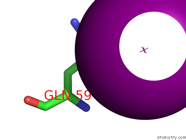
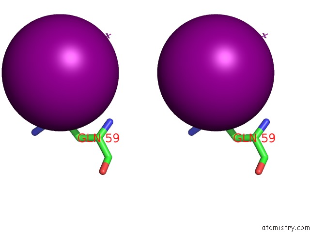
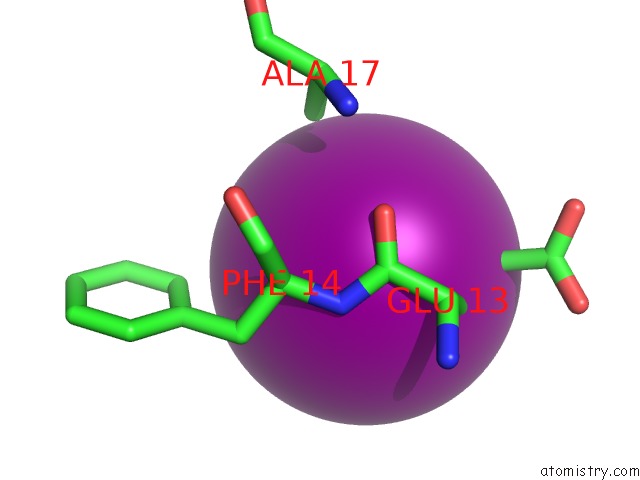
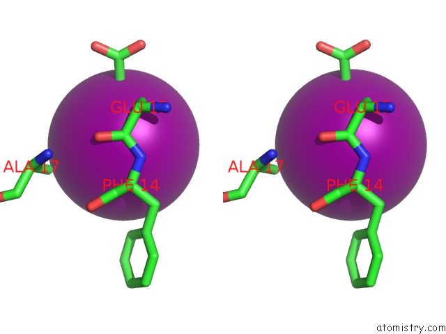
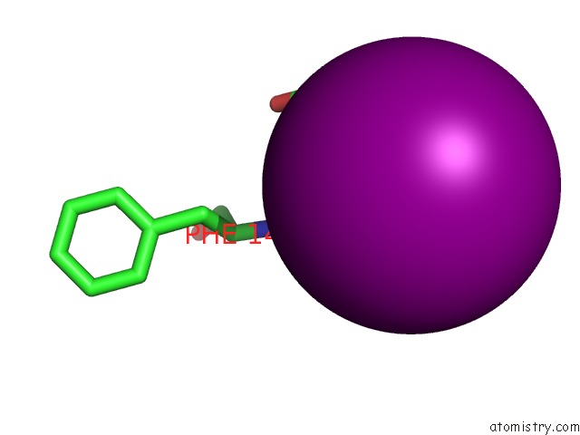
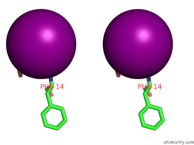
Iodine binding site 1 out of 7 in 3i7t
Go back to
Iodine binding site 1 out
of 7 in the Crystal Structure of RV2704, A Member of Highly Conserved Yjgf/YER057C/UK114 Family, From Mycobacterium Tuberculosis
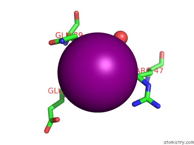
Mono view
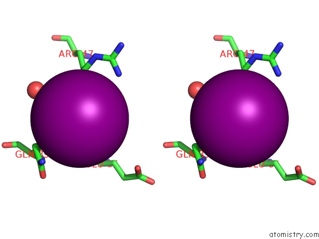
Stereo pair view
Mono view
Stereo pair view
A full contact list of Iodine with other atoms in the I binding
site number 1 of Crystal Structure of RV2704, A Member of Highly Conserved Yjgf/YER057C/UK114 Family, From Mycobacterium Tuberculosis within 5.0Å range:
|
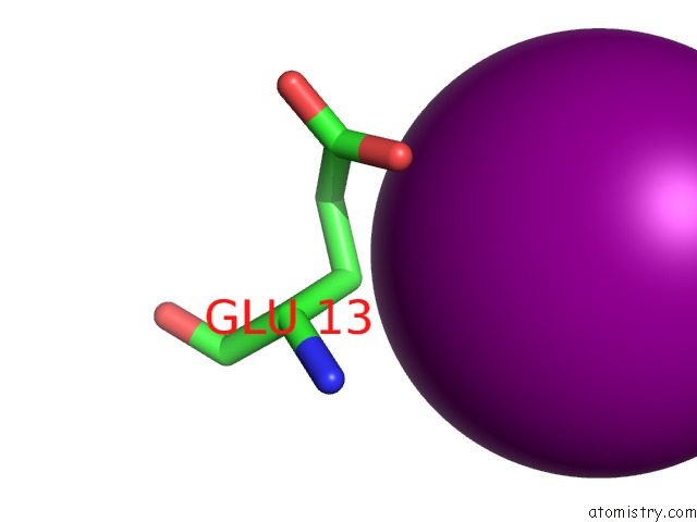
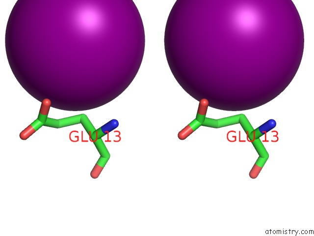
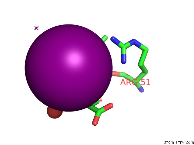
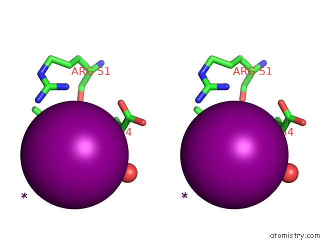
Iodine binding site 2 out of 7 in 3i7t
Go back to
Iodine binding site 2 out
of 7 in the Crystal Structure of RV2704, A Member of Highly Conserved Yjgf/YER057C/UK114 Family, From Mycobacterium Tuberculosis
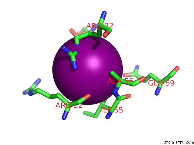
Mono view
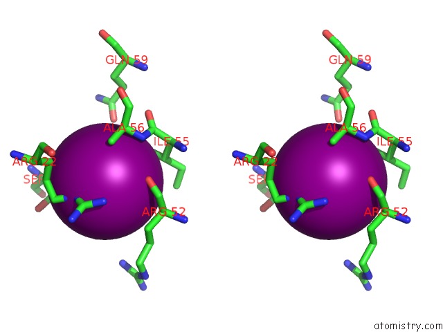
Stereo pair view
Mono view
Stereo pair view
A full contact list of Iodine with other atoms in the I binding
site number 2 of Crystal Structure of RV2704, A Member of Highly Conserved Yjgf/YER057C/UK114 Family, From Mycobacterium Tuberculosis within 5.0Å range:
|
Iodine binding site 3 out of 7 in 3i7t
Go back to
Iodine binding site 3 out
of 7 in the Crystal Structure of RV2704, A Member of Highly Conserved Yjgf/YER057C/UK114 Family, From Mycobacterium Tuberculosis
Mono view
Stereo pair view
Mono view
Stereo pair view
A full contact list of Iodine with other atoms in the I binding
site number 3 of Crystal Structure of RV2704, A Member of Highly Conserved Yjgf/YER057C/UK114 Family, From Mycobacterium Tuberculosis within 5.0Å range:
|
Iodine binding site 4 out of 7 in 3i7t
Go back to
Iodine binding site 4 out
of 7 in the Crystal Structure of RV2704, A Member of Highly Conserved Yjgf/YER057C/UK114 Family, From Mycobacterium Tuberculosis
Mono view
Stereo pair view
Mono view
Stereo pair view
A full contact list of Iodine with other atoms in the I binding
site number 4 of Crystal Structure of RV2704, A Member of Highly Conserved Yjgf/YER057C/UK114 Family, From Mycobacterium Tuberculosis within 5.0Å range:
|
Iodine binding site 5 out of 7 in 3i7t
Go back to
Iodine binding site 5 out
of 7 in the Crystal Structure of RV2704, A Member of Highly Conserved Yjgf/YER057C/UK114 Family, From Mycobacterium Tuberculosis
Mono view
Stereo pair view
Mono view
Stereo pair view
A full contact list of Iodine with other atoms in the I binding
site number 5 of Crystal Structure of RV2704, A Member of Highly Conserved Yjgf/YER057C/UK114 Family, From Mycobacterium Tuberculosis within 5.0Å range:
|
Iodine binding site 6 out of 7 in 3i7t
Go back to
Iodine binding site 6 out
of 7 in the Crystal Structure of RV2704, A Member of Highly Conserved Yjgf/YER057C/UK114 Family, From Mycobacterium Tuberculosis
Mono view
Stereo pair view
Mono view
Stereo pair view
A full contact list of Iodine with other atoms in the I binding
site number 6 of Crystal Structure of RV2704, A Member of Highly Conserved Yjgf/YER057C/UK114 Family, From Mycobacterium Tuberculosis within 5.0Å range:
|
Iodine binding site 7 out of 7 in 3i7t
Go back to
Iodine binding site 7 out
of 7 in the Crystal Structure of RV2704, A Member of Highly Conserved Yjgf/YER057C/UK114 Family, From Mycobacterium Tuberculosis
Mono view
Stereo pair view
Mono view
Stereo pair view
A full contact list of Iodine with other atoms in the I binding
site number 7 of Crystal Structure of RV2704, A Member of Highly Conserved Yjgf/YER057C/UK114 Family, From Mycobacterium Tuberculosis within 5.0Å range:
|
Reference:
K.G.Thakur,
T.Praveena,
B.Gopal.
Mycobacterium Tuberculosis RV2704 Is A Member of the Yjgf/YER057C/UK114 Family. Proteins V. 78 773 2009.
ISSN: ISSN 0887-3585
PubMed: 19899170
DOI: 10.1002/PROT.22623
Page generated: Sun Aug 11 15:21:06 2024
ISSN: ISSN 0887-3585
PubMed: 19899170
DOI: 10.1002/PROT.22623
Last articles
Zn in 9J0NZn in 9J0O
Zn in 9J0P
Zn in 9FJX
Zn in 9EKB
Zn in 9C0F
Zn in 9CAH
Zn in 9CH0
Zn in 9CH3
Zn in 9CH1