Iodine »
PDB 3kxn-3otd »
3mbl »
Iodine in PDB 3mbl: Crystal Structure of the Human Mitogen-Activated Protein Kinase Kinase 1 (Mek 1) in Complex with Ligand and Mgadp
Enzymatic activity of Crystal Structure of the Human Mitogen-Activated Protein Kinase Kinase 1 (Mek 1) in Complex with Ligand and Mgadp
All present enzymatic activity of Crystal Structure of the Human Mitogen-Activated Protein Kinase Kinase 1 (Mek 1) in Complex with Ligand and Mgadp:
2.7.12.2;
2.7.12.2;
Protein crystallography data
The structure of Crystal Structure of the Human Mitogen-Activated Protein Kinase Kinase 1 (Mek 1) in Complex with Ligand and Mgadp, PDB code: 3mbl
was solved by
D.R.Dougan,
C.D.Mol,
with X-Ray Crystallography technique. A brief refinement statistics is given in the table below:
| Resolution Low / High (Å) | 20.00 / 2.60 |
| Space group | P 62 |
| Cell size a, b, c (Å), α, β, γ (°) | 81.982, 81.982, 129.928, 90.00, 90.00, 120.00 |
| R / Rfree (%) | 16.6 / 21.5 |
Other elements in 3mbl:
The structure of Crystal Structure of the Human Mitogen-Activated Protein Kinase Kinase 1 (Mek 1) in Complex with Ligand and Mgadp also contains other interesting chemical elements:
| Fluorine | (F) | 1 atom |
| Magnesium | (Mg) | 1 atom |
Iodine Binding Sites:
The binding sites of Iodine atom in the Crystal Structure of the Human Mitogen-Activated Protein Kinase Kinase 1 (Mek 1) in Complex with Ligand and Mgadp
(pdb code 3mbl). This binding sites where shown within
5.0 Angstroms radius around Iodine atom.
In total only one binding site of Iodine was determined in the Crystal Structure of the Human Mitogen-Activated Protein Kinase Kinase 1 (Mek 1) in Complex with Ligand and Mgadp, PDB code: 3mbl:
In total only one binding site of Iodine was determined in the Crystal Structure of the Human Mitogen-Activated Protein Kinase Kinase 1 (Mek 1) in Complex with Ligand and Mgadp, PDB code: 3mbl:
Iodine binding site 1 out of 1 in 3mbl
Go back to
Iodine binding site 1 out
of 1 in the Crystal Structure of the Human Mitogen-Activated Protein Kinase Kinase 1 (Mek 1) in Complex with Ligand and Mgadp
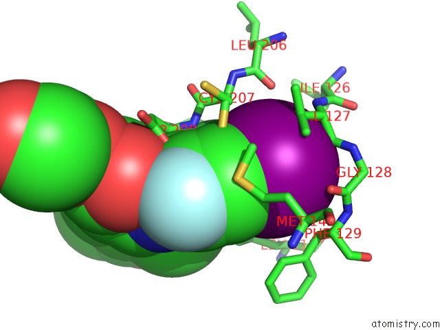
Mono view
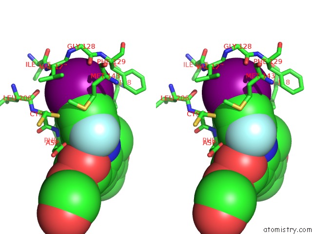
Stereo pair view
Mono view
Stereo pair view
A full contact list of Iodine with other atoms in the I binding
site number 1 of Crystal Structure of the Human Mitogen-Activated Protein Kinase Kinase 1 (Mek 1) in Complex with Ligand and Mgadp within 5.0Å range:
|
Reference:
M.B.Wallace,
M.E.Adams,
T.Kanouni,
C.D.Mol,
D.R.Dougan,
V.A.Feher,
S.M.O'connell,
L.Shi,
P.Halkowycz,
Q.Dong.
Structure-Based Design and Synthesis of Pyrrole Derivatives As Mek Inhibitors. Bioorg.Med.Chem.Lett. V. 20 4156 2010.
ISSN: ISSN 0960-894X
PubMed: 20621728
DOI: 10.1016/J.BMCL.2010.05.058
Page generated: Fri Aug 8 14:41:46 2025
ISSN: ISSN 0960-894X
PubMed: 20621728
DOI: 10.1016/J.BMCL.2010.05.058
Last articles
I in 8D22I in 8CXW
I in 8D00
I in 8CT8
I in 8CUF
I in 8CJ7
I in 8CNZ
I in 8C3P
I in 8CEV
I in 8C7R