Iodine »
PDB 3oth-3rtn »
3qs2 »
Iodine in PDB 3qs2: Crystal Structure of the Biofilm Forming Subunit of the E. Coli Common Pilus: Full Length Domain Swapped Dimer of Ecpa
Protein crystallography data
The structure of Crystal Structure of the Biofilm Forming Subunit of the E. Coli Common Pilus: Full Length Domain Swapped Dimer of Ecpa, PDB code: 3qs2
was solved by
J.A.Garnett,
S.J.Matthews,
with X-Ray Crystallography technique. A brief refinement statistics is given in the table below:
| Resolution Low / High (Å) | 60.01 / 1.78 |
| Space group | P 21 21 21 |
| Cell size a, b, c (Å), α, β, γ (°) | 46.860, 72.080, 108.320, 90.00, 90.00, 90.00 |
| R / Rfree (%) | 17.9 / 22 |
Iodine Binding Sites:
Pages:
>>> Page 1 <<< Page 2, Binding sites: 11 - 20; Page 3, Binding sites: 21 - 30; Page 4, Binding sites: 31 - 31;Binding sites:
The binding sites of Iodine atom in the Crystal Structure of the Biofilm Forming Subunit of the E. Coli Common Pilus: Full Length Domain Swapped Dimer of Ecpa (pdb code 3qs2). This binding sites where shown within 5.0 Angstroms radius around Iodine atom.In total 31 binding sites of Iodine where determined in the Crystal Structure of the Biofilm Forming Subunit of the E. Coli Common Pilus: Full Length Domain Swapped Dimer of Ecpa, PDB code: 3qs2:
Jump to Iodine binding site number: 1; 2; 3; 4; 5; 6; 7; 8; 9; 10;
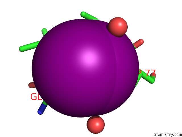
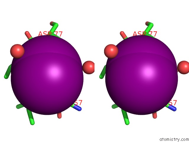
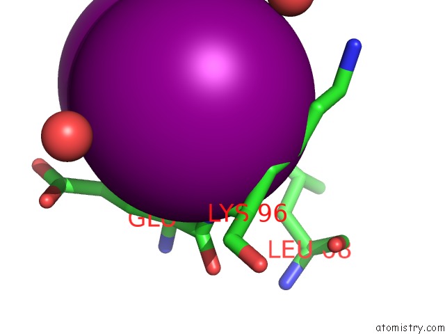
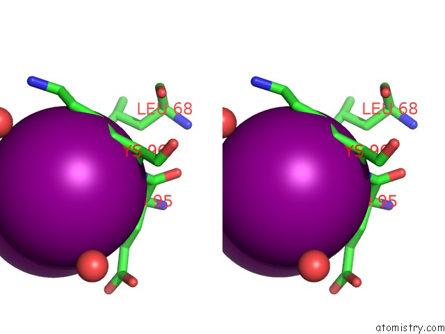
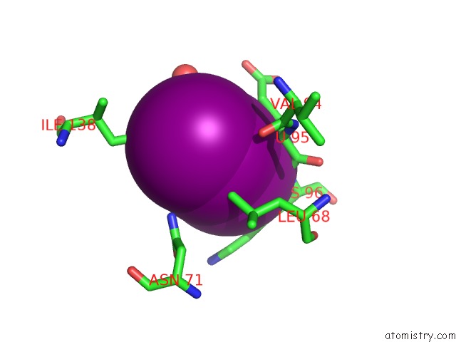
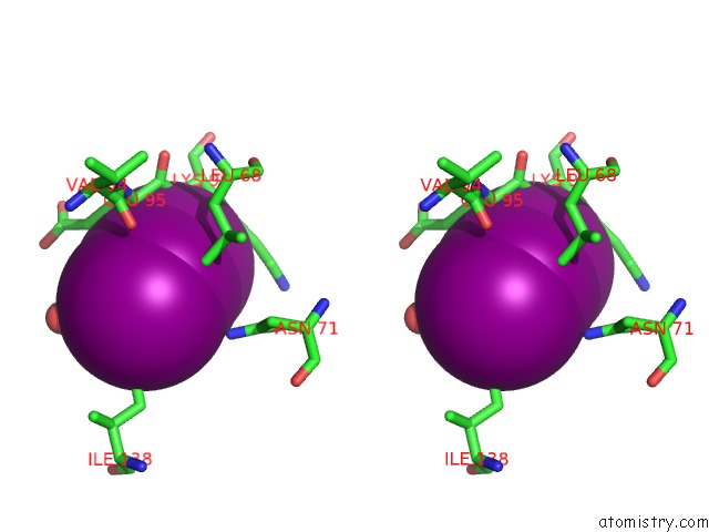
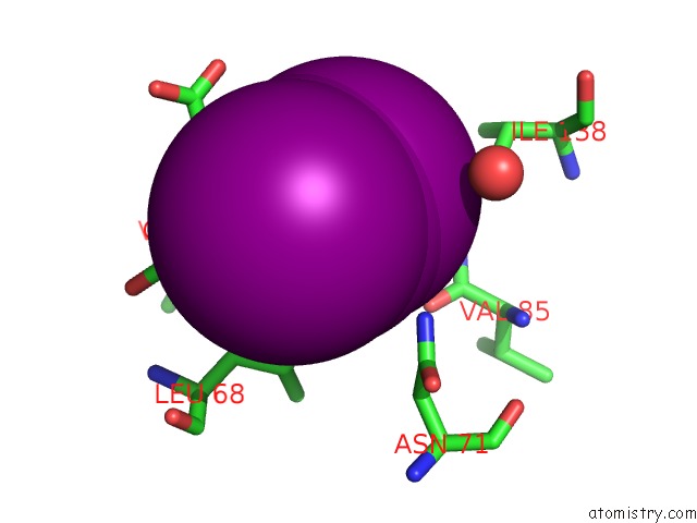
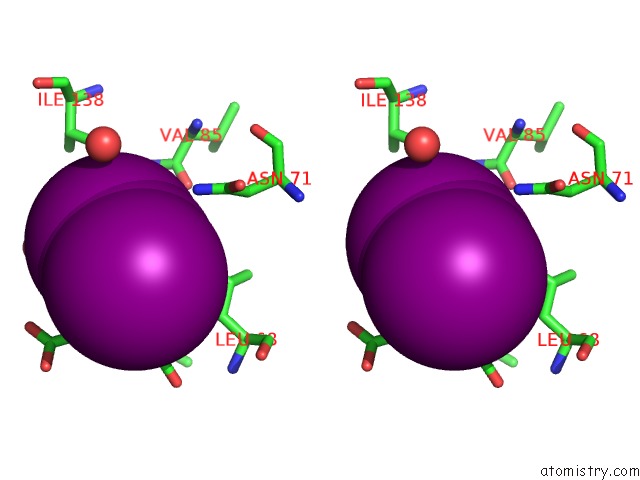
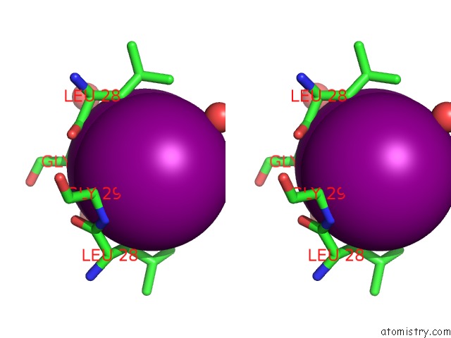
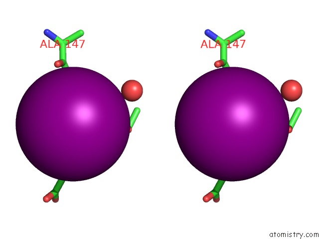
Iodine binding site 1 out of 31 in 3qs2
Go back to
Iodine binding site 1 out
of 31 in the Crystal Structure of the Biofilm Forming Subunit of the E. Coli Common Pilus: Full Length Domain Swapped Dimer of Ecpa
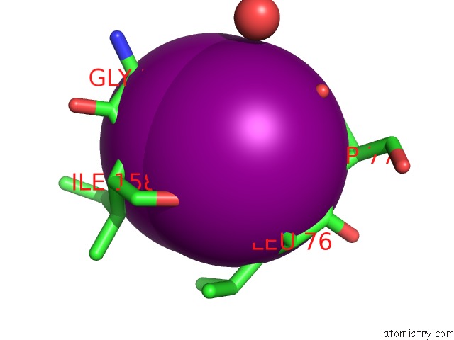
Mono view

Stereo pair view
Mono view
Stereo pair view
A full contact list of Iodine with other atoms in the I binding
site number 1 of Crystal Structure of the Biofilm Forming Subunit of the E. Coli Common Pilus: Full Length Domain Swapped Dimer of Ecpa within 5.0Å range:
|
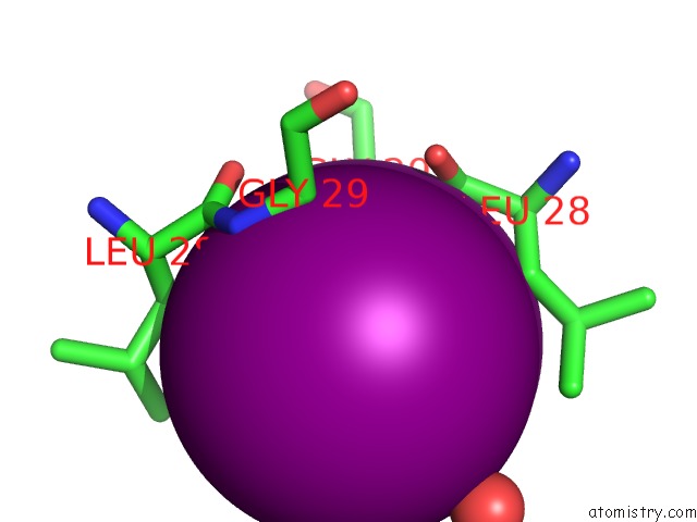
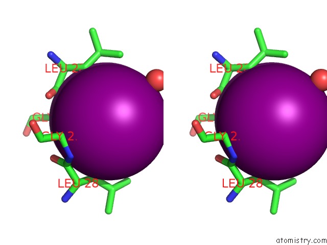
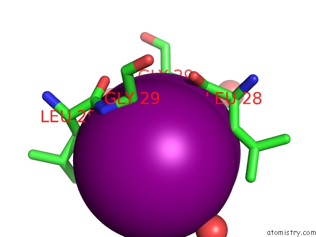
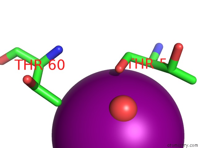
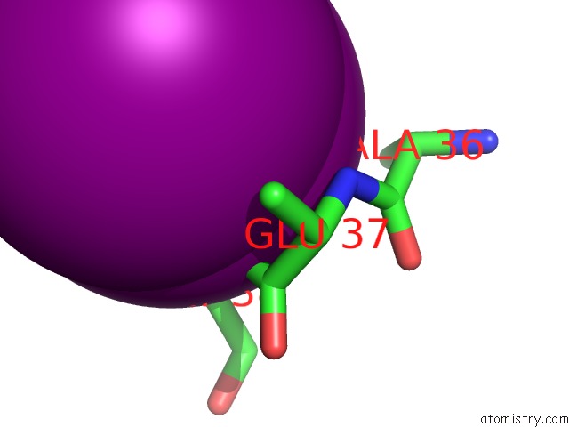
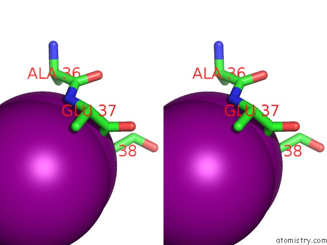
Iodine binding site 2 out of 31 in 3qs2
Go back to
Iodine binding site 2 out
of 31 in the Crystal Structure of the Biofilm Forming Subunit of the E. Coli Common Pilus: Full Length Domain Swapped Dimer of Ecpa
Mono view
Stereo pair view
Mono view
Stereo pair view
A full contact list of Iodine with other atoms in the I binding
site number 2 of Crystal Structure of the Biofilm Forming Subunit of the E. Coli Common Pilus: Full Length Domain Swapped Dimer of Ecpa within 5.0Å range:
|
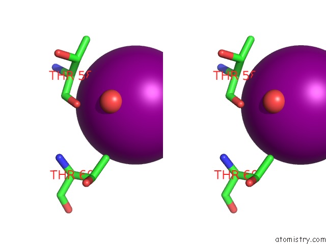
Iodine binding site 3 out of 31 in 3qs2
Go back to
Iodine binding site 3 out
of 31 in the Crystal Structure of the Biofilm Forming Subunit of the E. Coli Common Pilus: Full Length Domain Swapped Dimer of Ecpa
Mono view
Stereo pair view
Mono view
Stereo pair view
A full contact list of Iodine with other atoms in the I binding
site number 3 of Crystal Structure of the Biofilm Forming Subunit of the E. Coli Common Pilus: Full Length Domain Swapped Dimer of Ecpa within 5.0Å range:
|
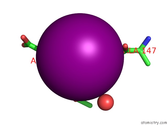
Iodine binding site 4 out of 31 in 3qs2
Go back to
Iodine binding site 4 out
of 31 in the Crystal Structure of the Biofilm Forming Subunit of the E. Coli Common Pilus: Full Length Domain Swapped Dimer of Ecpa
Mono view
Stereo pair view
Mono view
Stereo pair view
A full contact list of Iodine with other atoms in the I binding
site number 4 of Crystal Structure of the Biofilm Forming Subunit of the E. Coli Common Pilus: Full Length Domain Swapped Dimer of Ecpa within 5.0Å range:
|
Iodine binding site 5 out of 31 in 3qs2
Go back to
Iodine binding site 5 out
of 31 in the Crystal Structure of the Biofilm Forming Subunit of the E. Coli Common Pilus: Full Length Domain Swapped Dimer of Ecpa
Mono view
Stereo pair view
Mono view
Stereo pair view
A full contact list of Iodine with other atoms in the I binding
site number 5 of Crystal Structure of the Biofilm Forming Subunit of the E. Coli Common Pilus: Full Length Domain Swapped Dimer of Ecpa within 5.0Å range:
|
Iodine binding site 6 out of 31 in 3qs2
Go back to
Iodine binding site 6 out
of 31 in the Crystal Structure of the Biofilm Forming Subunit of the E. Coli Common Pilus: Full Length Domain Swapped Dimer of Ecpa
Mono view
Stereo pair view
Mono view
Stereo pair view
A full contact list of Iodine with other atoms in the I binding
site number 6 of Crystal Structure of the Biofilm Forming Subunit of the E. Coli Common Pilus: Full Length Domain Swapped Dimer of Ecpa within 5.0Å range:
|
Iodine binding site 7 out of 31 in 3qs2
Go back to
Iodine binding site 7 out
of 31 in the Crystal Structure of the Biofilm Forming Subunit of the E. Coli Common Pilus: Full Length Domain Swapped Dimer of Ecpa
Mono view
Stereo pair view
Mono view
Stereo pair view
A full contact list of Iodine with other atoms in the I binding
site number 7 of Crystal Structure of the Biofilm Forming Subunit of the E. Coli Common Pilus: Full Length Domain Swapped Dimer of Ecpa within 5.0Å range:
|
Iodine binding site 8 out of 31 in 3qs2
Go back to
Iodine binding site 8 out
of 31 in the Crystal Structure of the Biofilm Forming Subunit of the E. Coli Common Pilus: Full Length Domain Swapped Dimer of Ecpa
Mono view
Stereo pair view
Mono view
Stereo pair view
A full contact list of Iodine with other atoms in the I binding
site number 8 of Crystal Structure of the Biofilm Forming Subunit of the E. Coli Common Pilus: Full Length Domain Swapped Dimer of Ecpa within 5.0Å range:
|
Iodine binding site 9 out of 31 in 3qs2
Go back to
Iodine binding site 9 out
of 31 in the Crystal Structure of the Biofilm Forming Subunit of the E. Coli Common Pilus: Full Length Domain Swapped Dimer of Ecpa
Mono view
Stereo pair view
Mono view
Stereo pair view
A full contact list of Iodine with other atoms in the I binding
site number 9 of Crystal Structure of the Biofilm Forming Subunit of the E. Coli Common Pilus: Full Length Domain Swapped Dimer of Ecpa within 5.0Å range:
|
Iodine binding site 10 out of 31 in 3qs2
Go back to
Iodine binding site 10 out
of 31 in the Crystal Structure of the Biofilm Forming Subunit of the E. Coli Common Pilus: Full Length Domain Swapped Dimer of Ecpa
Mono view
Stereo pair view
Mono view
Stereo pair view
A full contact list of Iodine with other atoms in the I binding
site number 10 of Crystal Structure of the Biofilm Forming Subunit of the E. Coli Common Pilus: Full Length Domain Swapped Dimer of Ecpa within 5.0Å range:
|
Reference:
J.A.Garnett,
V.I.Martinez-Santos,
Z.Saldana,
T.Pape,
W.Hawthorne,
J.Chan,
P.J.Simpson,
E.Cota,
J.L.Puente,
J.A.Giron,
S.Matthews.
Structural Insights Into the Biogenesis and Biofilm Formation By the Escherichia Coli Common Pilus. Proc.Natl.Acad.Sci.Usa V. 109 3950 2012.
ISSN: ISSN 0027-8424
PubMed: 22355107
DOI: 10.1073/PNAS.1106733109
Page generated: Sun Aug 11 16:17:01 2024
ISSN: ISSN 0027-8424
PubMed: 22355107
DOI: 10.1073/PNAS.1106733109
Last articles
Zn in 9J0NZn in 9J0O
Zn in 9J0P
Zn in 9FJX
Zn in 9EKB
Zn in 9C0F
Zn in 9CAH
Zn in 9CH0
Zn in 9CH3
Zn in 9CH1