Iodine »
PDB 3oth-3rtn »
3r2v »
Iodine in PDB 3r2v: Crystal Structure of Polymerase Basic Protein 2 E538-R753 From Influenza A Virus A/Yokohama/2017/03 H3N2
Protein crystallography data
The structure of Crystal Structure of Polymerase Basic Protein 2 E538-R753 From Influenza A Virus A/Yokohama/2017/03 H3N2, PDB code: 3r2v
was solved by
Seattle Structural Genomics Center For Infectious Disease (Ssgcid),
with X-Ray Crystallography technique. A brief refinement statistics is given in the table below:
| Resolution Low / High (Å) | 30.00 / 1.30 |
| Space group | P 21 21 2 |
| Cell size a, b, c (Å), α, β, γ (°) | 52.810, 106.540, 33.490, 90.00, 90.00, 90.00 |
| R / Rfree (%) | 18 / 20.9 |
Iodine Binding Sites:
Pages:
>>> Page 1 <<< Page 2, Binding sites: 11 - 12;Binding sites:
The binding sites of Iodine atom in the Crystal Structure of Polymerase Basic Protein 2 E538-R753 From Influenza A Virus A/Yokohama/2017/03 H3N2 (pdb code 3r2v). This binding sites where shown within 5.0 Angstroms radius around Iodine atom.In total 12 binding sites of Iodine where determined in the Crystal Structure of Polymerase Basic Protein 2 E538-R753 From Influenza A Virus A/Yokohama/2017/03 H3N2, PDB code: 3r2v:
Jump to Iodine binding site number: 1; 2; 3; 4; 5; 6; 7; 8; 9; 10;
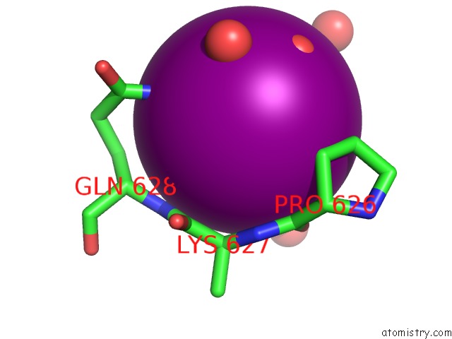
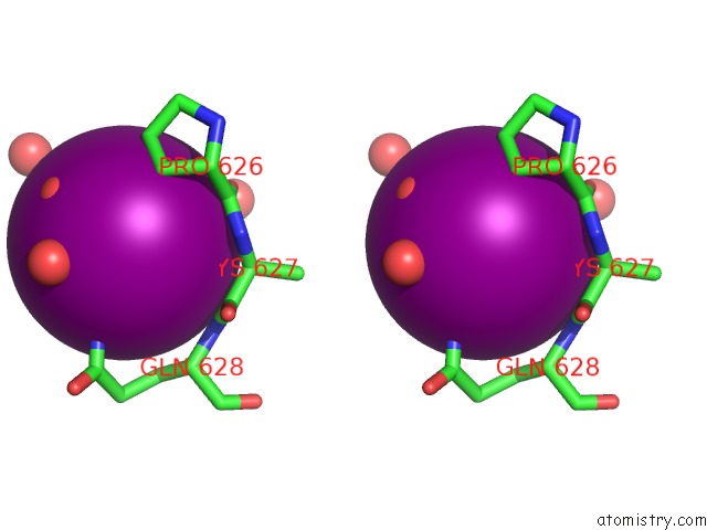
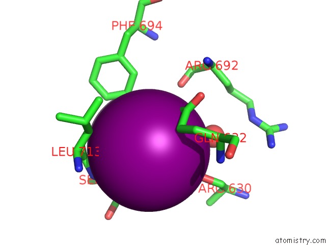
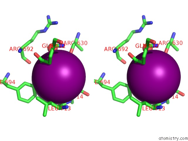
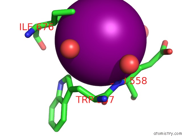
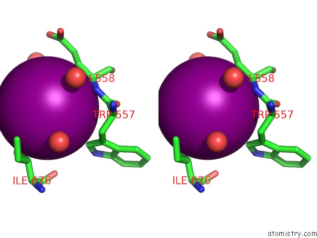
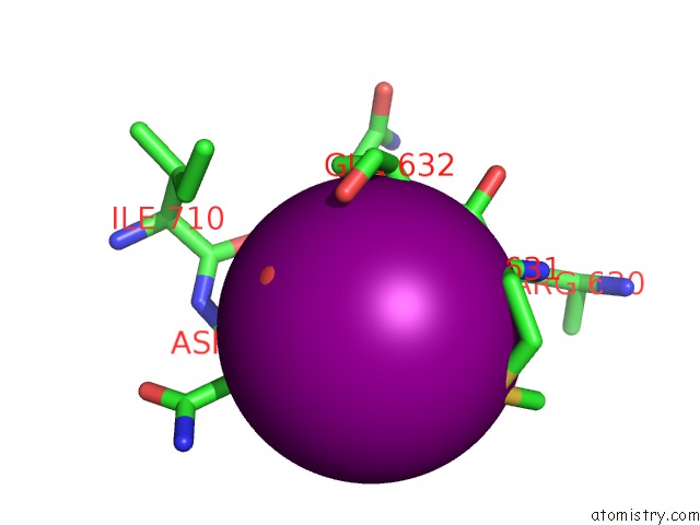
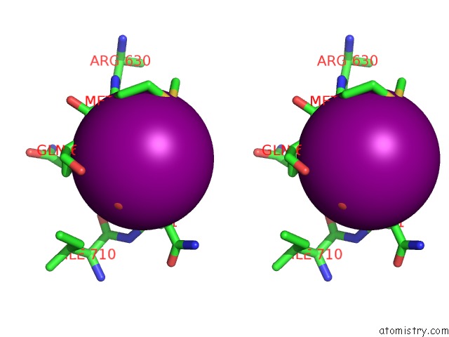
Iodine binding site 1 out of 12 in 3r2v
Go back to
Iodine binding site 1 out
of 12 in the Crystal Structure of Polymerase Basic Protein 2 E538-R753 From Influenza A Virus A/Yokohama/2017/03 H3N2
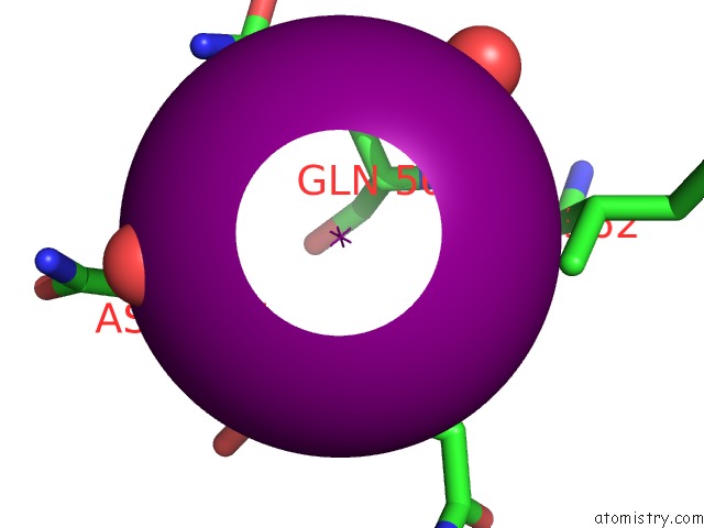
Mono view
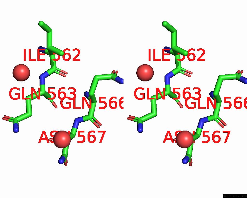
Stereo pair view
Mono view
Stereo pair view
A full contact list of Iodine with other atoms in the I binding
site number 1 of Crystal Structure of Polymerase Basic Protein 2 E538-R753 From Influenza A Virus A/Yokohama/2017/03 H3N2 within 5.0Å range:
|
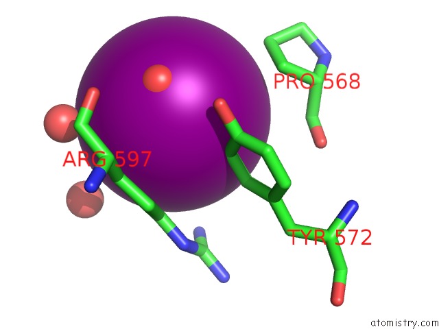
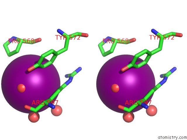
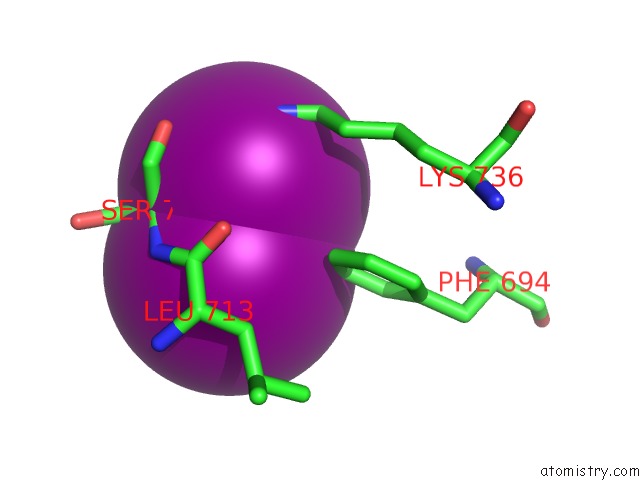
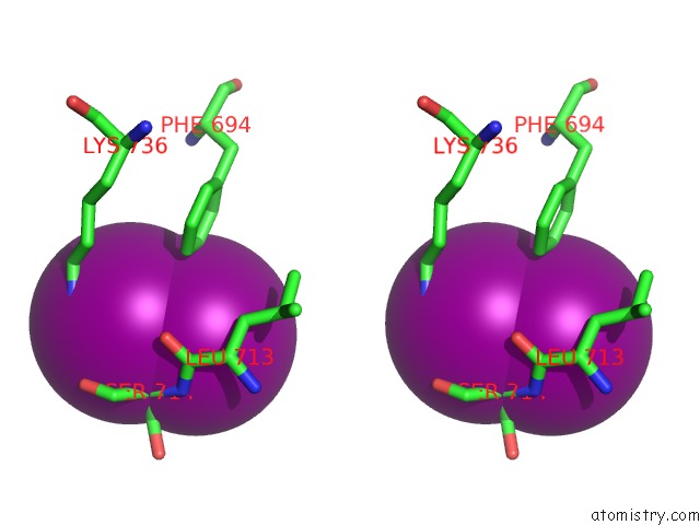
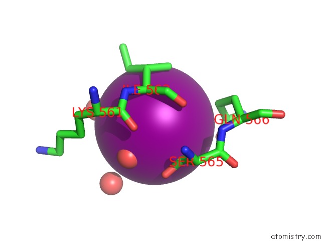
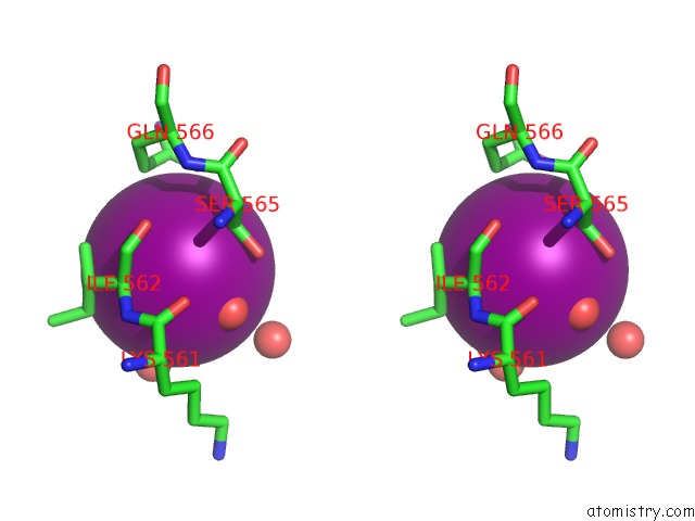
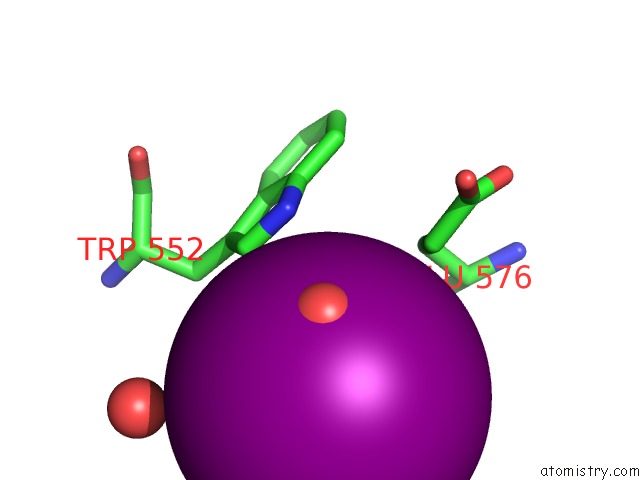
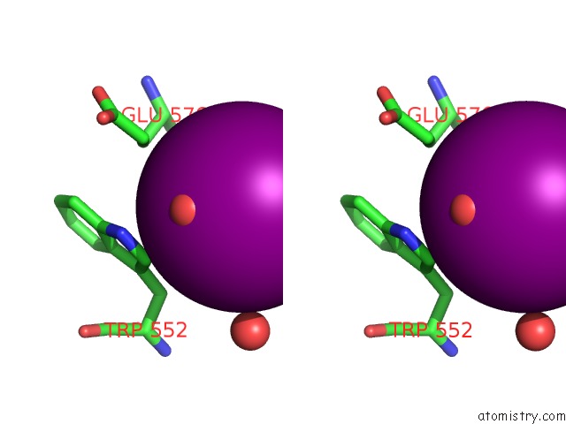
Iodine binding site 2 out of 12 in 3r2v
Go back to
Iodine binding site 2 out
of 12 in the Crystal Structure of Polymerase Basic Protein 2 E538-R753 From Influenza A Virus A/Yokohama/2017/03 H3N2
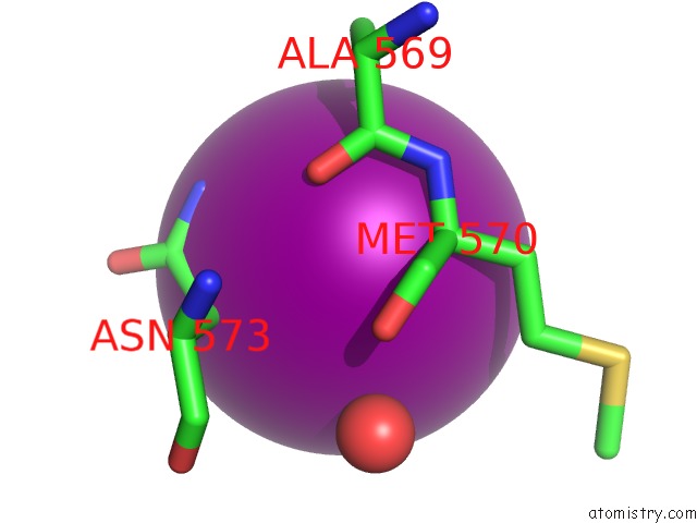
Mono view
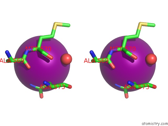
Stereo pair view
Mono view
Stereo pair view
A full contact list of Iodine with other atoms in the I binding
site number 2 of Crystal Structure of Polymerase Basic Protein 2 E538-R753 From Influenza A Virus A/Yokohama/2017/03 H3N2 within 5.0Å range:
|
Iodine binding site 3 out of 12 in 3r2v
Go back to
Iodine binding site 3 out
of 12 in the Crystal Structure of Polymerase Basic Protein 2 E538-R753 From Influenza A Virus A/Yokohama/2017/03 H3N2
Mono view
Stereo pair view
Mono view
Stereo pair view
A full contact list of Iodine with other atoms in the I binding
site number 3 of Crystal Structure of Polymerase Basic Protein 2 E538-R753 From Influenza A Virus A/Yokohama/2017/03 H3N2 within 5.0Å range:
|
Iodine binding site 4 out of 12 in 3r2v
Go back to
Iodine binding site 4 out
of 12 in the Crystal Structure of Polymerase Basic Protein 2 E538-R753 From Influenza A Virus A/Yokohama/2017/03 H3N2
Mono view
Stereo pair view
Mono view
Stereo pair view
A full contact list of Iodine with other atoms in the I binding
site number 4 of Crystal Structure of Polymerase Basic Protein 2 E538-R753 From Influenza A Virus A/Yokohama/2017/03 H3N2 within 5.0Å range:
|
Iodine binding site 5 out of 12 in 3r2v
Go back to
Iodine binding site 5 out
of 12 in the Crystal Structure of Polymerase Basic Protein 2 E538-R753 From Influenza A Virus A/Yokohama/2017/03 H3N2
Mono view
Stereo pair view
Mono view
Stereo pair view
A full contact list of Iodine with other atoms in the I binding
site number 5 of Crystal Structure of Polymerase Basic Protein 2 E538-R753 From Influenza A Virus A/Yokohama/2017/03 H3N2 within 5.0Å range:
|
Iodine binding site 6 out of 12 in 3r2v
Go back to
Iodine binding site 6 out
of 12 in the Crystal Structure of Polymerase Basic Protein 2 E538-R753 From Influenza A Virus A/Yokohama/2017/03 H3N2
Mono view
Stereo pair view
Mono view
Stereo pair view
A full contact list of Iodine with other atoms in the I binding
site number 6 of Crystal Structure of Polymerase Basic Protein 2 E538-R753 From Influenza A Virus A/Yokohama/2017/03 H3N2 within 5.0Å range:
|
Iodine binding site 7 out of 12 in 3r2v
Go back to
Iodine binding site 7 out
of 12 in the Crystal Structure of Polymerase Basic Protein 2 E538-R753 From Influenza A Virus A/Yokohama/2017/03 H3N2
Mono view
Stereo pair view
Mono view
Stereo pair view
A full contact list of Iodine with other atoms in the I binding
site number 7 of Crystal Structure of Polymerase Basic Protein 2 E538-R753 From Influenza A Virus A/Yokohama/2017/03 H3N2 within 5.0Å range:
|
Iodine binding site 8 out of 12 in 3r2v
Go back to
Iodine binding site 8 out
of 12 in the Crystal Structure of Polymerase Basic Protein 2 E538-R753 From Influenza A Virus A/Yokohama/2017/03 H3N2
Mono view
Stereo pair view
Mono view
Stereo pair view
A full contact list of Iodine with other atoms in the I binding
site number 8 of Crystal Structure of Polymerase Basic Protein 2 E538-R753 From Influenza A Virus A/Yokohama/2017/03 H3N2 within 5.0Å range:
|
Iodine binding site 9 out of 12 in 3r2v
Go back to
Iodine binding site 9 out
of 12 in the Crystal Structure of Polymerase Basic Protein 2 E538-R753 From Influenza A Virus A/Yokohama/2017/03 H3N2
Mono view
Stereo pair view
Mono view
Stereo pair view
A full contact list of Iodine with other atoms in the I binding
site number 9 of Crystal Structure of Polymerase Basic Protein 2 E538-R753 From Influenza A Virus A/Yokohama/2017/03 H3N2 within 5.0Å range:
|
Iodine binding site 10 out of 12 in 3r2v
Go back to
Iodine binding site 10 out
of 12 in the Crystal Structure of Polymerase Basic Protein 2 E538-R753 From Influenza A Virus A/Yokohama/2017/03 H3N2
Mono view
Stereo pair view
Mono view
Stereo pair view
A full contact list of Iodine with other atoms in the I binding
site number 10 of Crystal Structure of Polymerase Basic Protein 2 E538-R753 From Influenza A Virus A/Yokohama/2017/03 H3N2 within 5.0Å range:
|
Reference:
E.R.Smith,
D.W.Begley,
V.Anderson,
A.C.Raymond,
T.E.Haffner,
J.I.Robinson,
T.E.Edwards,
N.Duncan,
C.J.Gerdts,
M.B.Mixon,
P.Nollert,
B.L.Staker,
L.J.Stewart.
The Protein Maker: An Automated System For High-Throughput Parallel Purification. Acta Crystallogr.,Sect.F V. 67 1015 2011.
ISSN: ESSN 1744-3091
PubMed: 21904043
DOI: 10.1107/S1744309111028776
Page generated: Sun Aug 11 16:17:17 2024
ISSN: ESSN 1744-3091
PubMed: 21904043
DOI: 10.1107/S1744309111028776
Last articles
Zn in 9J0NZn in 9J0O
Zn in 9J0P
Zn in 9FJX
Zn in 9EKB
Zn in 9C0F
Zn in 9CAH
Zn in 9CH0
Zn in 9CH3
Zn in 9CH1