Iodine »
PDB 3ru1-3unh »
3t96 »
Iodine in PDB 3t96: Iodowillardiine Bound to A Double Cysteine Mutant (A452C/S652C) of the Ligand Binding Domain of GLUA2
Protein crystallography data
The structure of Iodowillardiine Bound to A Double Cysteine Mutant (A452C/S652C) of the Ligand Binding Domain of GLUA2, PDB code: 3t96
was solved by
A.H.Ahmed,
S.Wang,
H.H.Chuang,
R.E.Oswald,
with X-Ray Crystallography technique. A brief refinement statistics is given in the table below:
| Resolution Low / High (Å) | 26.99 / 1.87 |
| Space group | P 2 21 21 |
| Cell size a, b, c (Å), α, β, γ (°) | 47.793, 114.351, 163.547, 90.00, 90.00, 90.00 |
| R / Rfree (%) | 18.3 / 21.9 |
Other elements in 3t96:
The structure of Iodowillardiine Bound to A Double Cysteine Mutant (A452C/S652C) of the Ligand Binding Domain of GLUA2 also contains other interesting chemical elements:
| Zinc | (Zn) | 5 atoms |
Iodine Binding Sites:
The binding sites of Iodine atom in the Iodowillardiine Bound to A Double Cysteine Mutant (A452C/S652C) of the Ligand Binding Domain of GLUA2
(pdb code 3t96). This binding sites where shown within
5.0 Angstroms radius around Iodine atom.
In total 3 binding sites of Iodine where determined in the Iodowillardiine Bound to A Double Cysteine Mutant (A452C/S652C) of the Ligand Binding Domain of GLUA2, PDB code: 3t96:
Jump to Iodine binding site number: 1; 2; 3;
In total 3 binding sites of Iodine where determined in the Iodowillardiine Bound to A Double Cysteine Mutant (A452C/S652C) of the Ligand Binding Domain of GLUA2, PDB code: 3t96:
Jump to Iodine binding site number: 1; 2; 3;
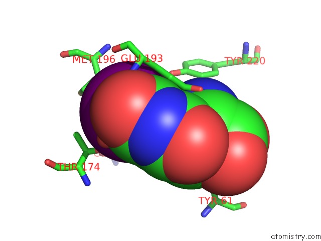
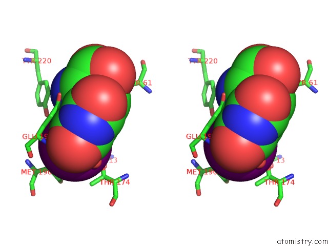
Iodine binding site 1 out of 3 in 3t96
Go back to
Iodine binding site 1 out
of 3 in the Iodowillardiine Bound to A Double Cysteine Mutant (A452C/S652C) of the Ligand Binding Domain of GLUA2
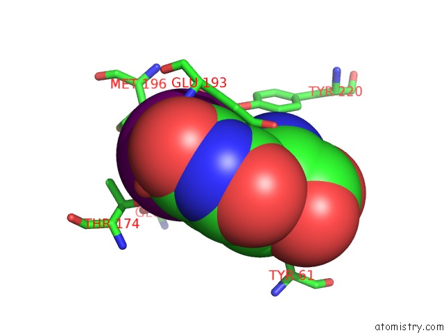
Mono view
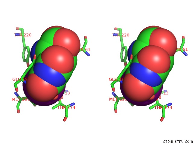
Stereo pair view
Mono view
Stereo pair view
A full contact list of Iodine with other atoms in the I binding
site number 1 of Iodowillardiine Bound to A Double Cysteine Mutant (A452C/S652C) of the Ligand Binding Domain of GLUA2 within 5.0Å range:
|
Iodine binding site 2 out of 3 in 3t96
Go back to
Iodine binding site 2 out
of 3 in the Iodowillardiine Bound to A Double Cysteine Mutant (A452C/S652C) of the Ligand Binding Domain of GLUA2
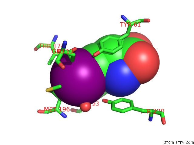
Mono view
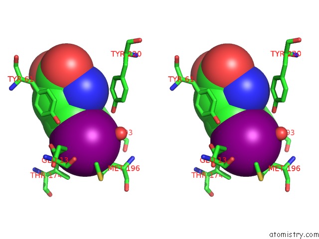
Stereo pair view
Mono view
Stereo pair view
A full contact list of Iodine with other atoms in the I binding
site number 2 of Iodowillardiine Bound to A Double Cysteine Mutant (A452C/S652C) of the Ligand Binding Domain of GLUA2 within 5.0Å range:
|
Iodine binding site 3 out of 3 in 3t96
Go back to
Iodine binding site 3 out
of 3 in the Iodowillardiine Bound to A Double Cysteine Mutant (A452C/S652C) of the Ligand Binding Domain of GLUA2
Mono view
Stereo pair view
Mono view
Stereo pair view
A full contact list of Iodine with other atoms in the I binding
site number 3 of Iodowillardiine Bound to A Double Cysteine Mutant (A452C/S652C) of the Ligand Binding Domain of GLUA2 within 5.0Å range:
|
Reference:
A.H.Ahmed,
S.Wang,
H.H.Chuang,
R.E.Oswald.
Mechanism of Ampa Receptor Activation By Partial Agonists: Disulfide Trapping of Closed Lobe Conformations. J.Biol.Chem. V. 286 35257 2011.
ISSN: ISSN 0021-9258
PubMed: 21846932
DOI: 10.1074/JBC.M111.269001
Page generated: Sun Aug 11 16:50:31 2024
ISSN: ISSN 0021-9258
PubMed: 21846932
DOI: 10.1074/JBC.M111.269001
Last articles
Zn in 9MJ5Zn in 9HNW
Zn in 9G0L
Zn in 9FNE
Zn in 9DZN
Zn in 9E0I
Zn in 9D32
Zn in 9DAK
Zn in 8ZXC
Zn in 8ZUF