Iodine »
PDB 3ru1-3unh »
3tur »
Iodine in PDB 3tur: Crystal Structure of M. Tuberculosis Ld-Transpeptidase Type 2 Complexed with A Peptidoglycan Fragment
Protein crystallography data
The structure of Crystal Structure of M. Tuberculosis Ld-Transpeptidase Type 2 Complexed with A Peptidoglycan Fragment, PDB code: 3tur
was solved by
M.A.Bianchet,
S.B.Erdemli,
R.Gupta,
G.Lamichhane,
L.M.Amzel,
with X-Ray Crystallography technique. A brief refinement statistics is given in the table below:
| Resolution Low / High (Å) | 85.69 / 1.72 |
| Space group | I 21 21 21 |
| Cell size a, b, c (Å), α, β, γ (°) | 119.132, 120.829, 122.847, 90.00, 90.00, 90.00 |
| R / Rfree (%) | 19.9 / 23.5 |
Other elements in 3tur:
The structure of Crystal Structure of M. Tuberculosis Ld-Transpeptidase Type 2 Complexed with A Peptidoglycan Fragment also contains other interesting chemical elements:
| Platinum | (Pt) | 12 atoms |
Iodine Binding Sites:
Pages:
>>> Page 1 <<< Page 2, Binding sites: 11 - 11;Binding sites:
The binding sites of Iodine atom in the Crystal Structure of M. Tuberculosis Ld-Transpeptidase Type 2 Complexed with A Peptidoglycan Fragment (pdb code 3tur). This binding sites where shown within 5.0 Angstroms radius around Iodine atom.In total 11 binding sites of Iodine where determined in the Crystal Structure of M. Tuberculosis Ld-Transpeptidase Type 2 Complexed with A Peptidoglycan Fragment, PDB code: 3tur:
Jump to Iodine binding site number: 1; 2; 3; 4; 5; 6; 7; 8; 9; 10;
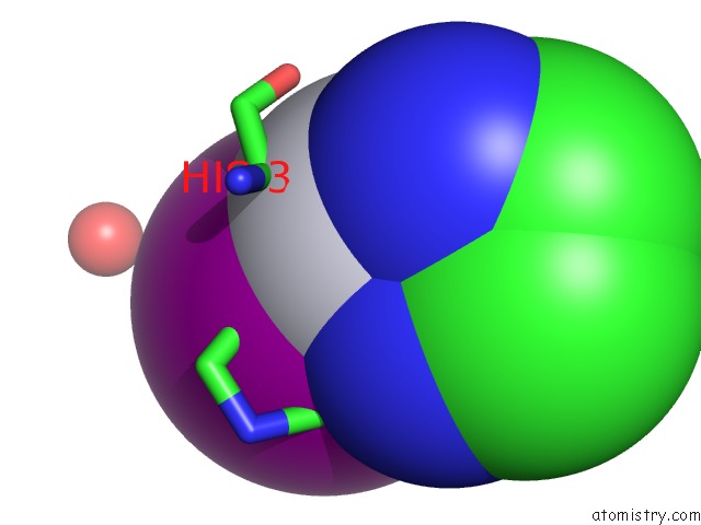
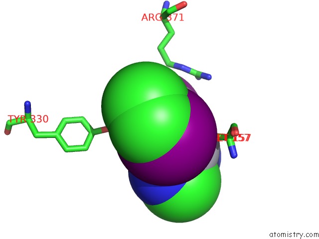
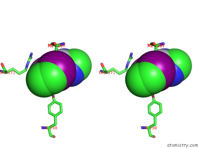
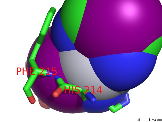
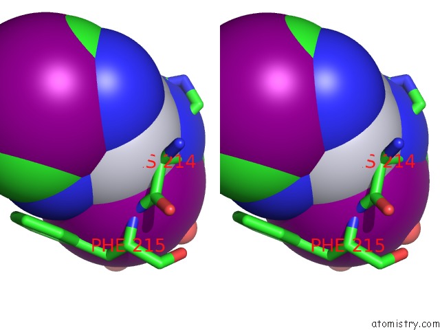
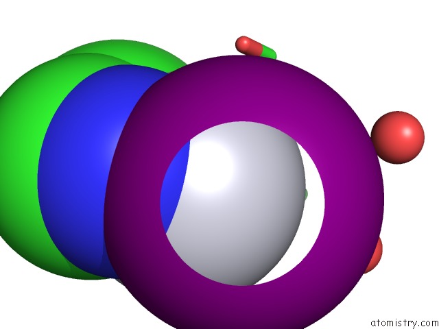
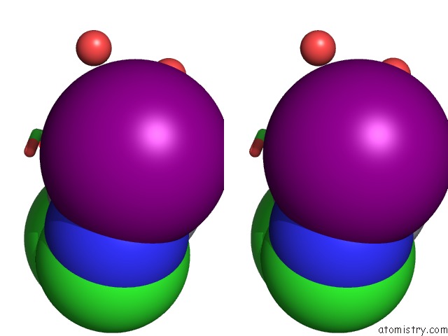
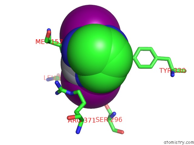
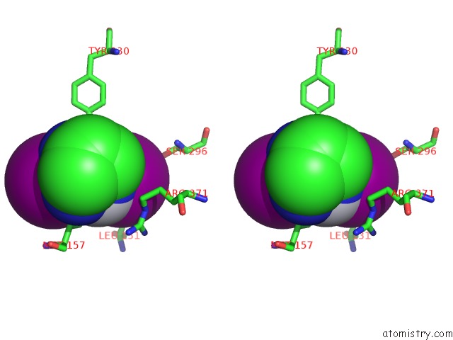
Iodine binding site 1 out of 11 in 3tur
Go back to
Iodine binding site 1 out
of 11 in the Crystal Structure of M. Tuberculosis Ld-Transpeptidase Type 2 Complexed with A Peptidoglycan Fragment
Mono view
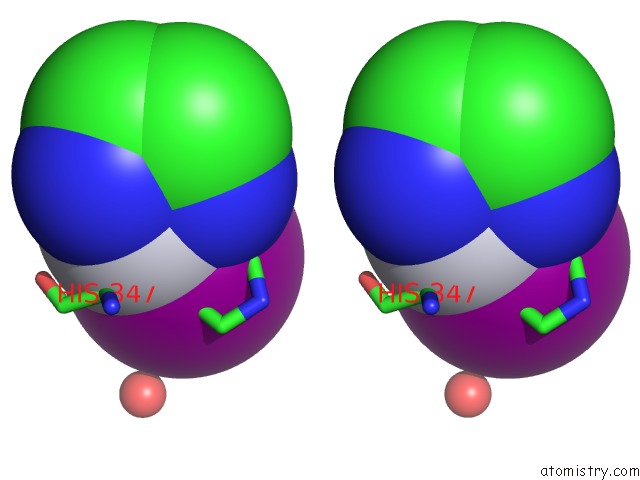
Stereo pair view
Mono view
Stereo pair view
A full contact list of Iodine with other atoms in the I binding
site number 1 of Crystal Structure of M. Tuberculosis Ld-Transpeptidase Type 2 Complexed with A Peptidoglycan Fragment within 5.0Å range:
|
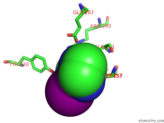
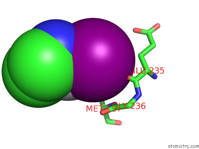
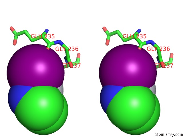
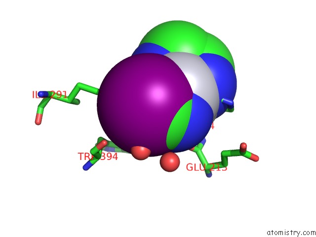
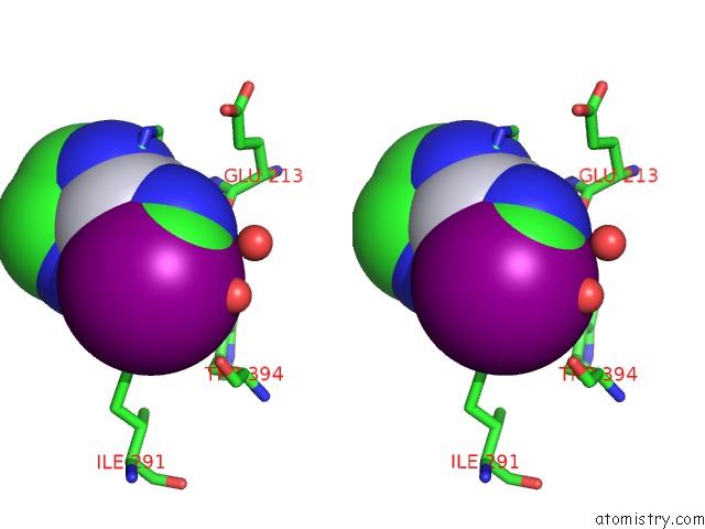
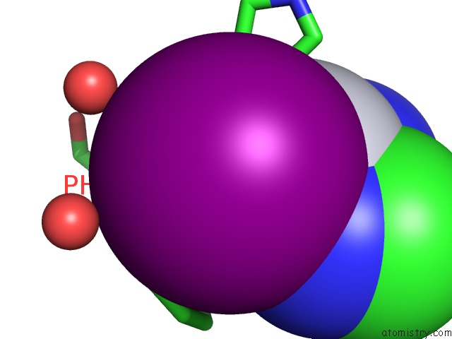
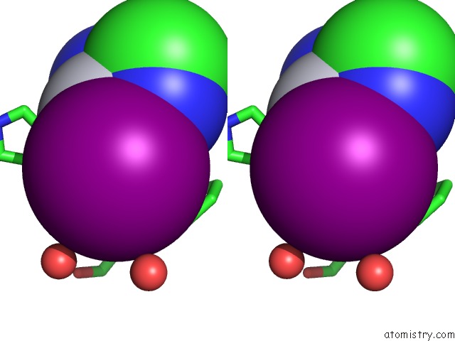
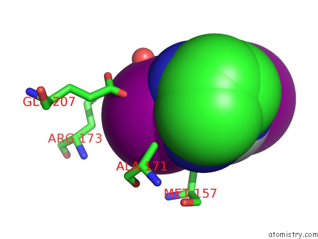
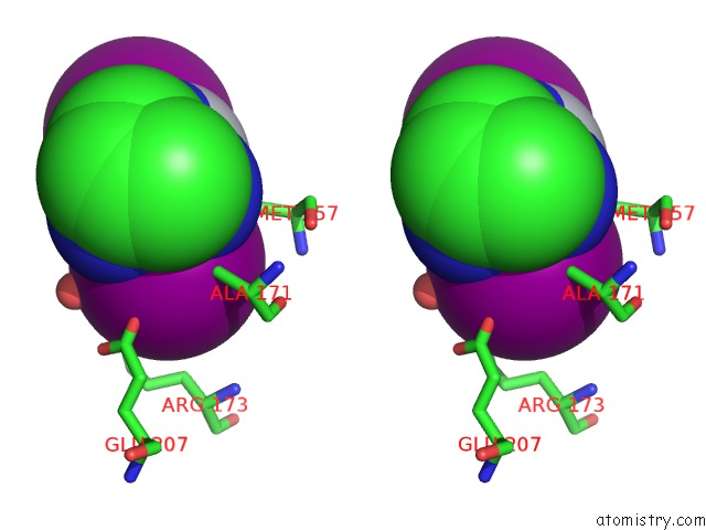
Iodine binding site 2 out of 11 in 3tur
Go back to
Iodine binding site 2 out
of 11 in the Crystal Structure of M. Tuberculosis Ld-Transpeptidase Type 2 Complexed with A Peptidoglycan Fragment
Mono view
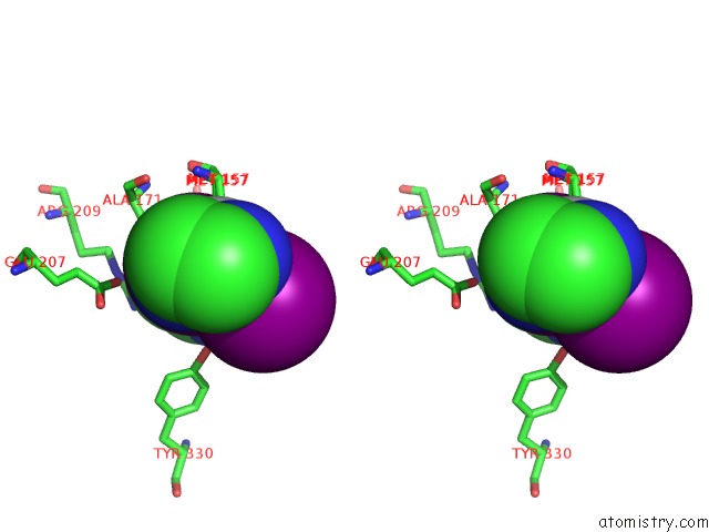
Stereo pair view
Mono view
Stereo pair view
A full contact list of Iodine with other atoms in the I binding
site number 2 of Crystal Structure of M. Tuberculosis Ld-Transpeptidase Type 2 Complexed with A Peptidoglycan Fragment within 5.0Å range:
|
Iodine binding site 3 out of 11 in 3tur
Go back to
Iodine binding site 3 out
of 11 in the Crystal Structure of M. Tuberculosis Ld-Transpeptidase Type 2 Complexed with A Peptidoglycan Fragment
Mono view
Stereo pair view
Mono view
Stereo pair view
A full contact list of Iodine with other atoms in the I binding
site number 3 of Crystal Structure of M. Tuberculosis Ld-Transpeptidase Type 2 Complexed with A Peptidoglycan Fragment within 5.0Å range:
|
Iodine binding site 4 out of 11 in 3tur
Go back to
Iodine binding site 4 out
of 11 in the Crystal Structure of M. Tuberculosis Ld-Transpeptidase Type 2 Complexed with A Peptidoglycan Fragment
Mono view
Stereo pair view
Mono view
Stereo pair view
A full contact list of Iodine with other atoms in the I binding
site number 4 of Crystal Structure of M. Tuberculosis Ld-Transpeptidase Type 2 Complexed with A Peptidoglycan Fragment within 5.0Å range:
|
Iodine binding site 5 out of 11 in 3tur
Go back to
Iodine binding site 5 out
of 11 in the Crystal Structure of M. Tuberculosis Ld-Transpeptidase Type 2 Complexed with A Peptidoglycan Fragment
Mono view
Stereo pair view
Mono view
Stereo pair view
A full contact list of Iodine with other atoms in the I binding
site number 5 of Crystal Structure of M. Tuberculosis Ld-Transpeptidase Type 2 Complexed with A Peptidoglycan Fragment within 5.0Å range:
|
Iodine binding site 6 out of 11 in 3tur
Go back to
Iodine binding site 6 out
of 11 in the Crystal Structure of M. Tuberculosis Ld-Transpeptidase Type 2 Complexed with A Peptidoglycan Fragment
Mono view
Stereo pair view
Mono view
Stereo pair view
A full contact list of Iodine with other atoms in the I binding
site number 6 of Crystal Structure of M. Tuberculosis Ld-Transpeptidase Type 2 Complexed with A Peptidoglycan Fragment within 5.0Å range:
|
Iodine binding site 7 out of 11 in 3tur
Go back to
Iodine binding site 7 out
of 11 in the Crystal Structure of M. Tuberculosis Ld-Transpeptidase Type 2 Complexed with A Peptidoglycan Fragment
Mono view
Stereo pair view
Mono view
Stereo pair view
A full contact list of Iodine with other atoms in the I binding
site number 7 of Crystal Structure of M. Tuberculosis Ld-Transpeptidase Type 2 Complexed with A Peptidoglycan Fragment within 5.0Å range:
|
Iodine binding site 8 out of 11 in 3tur
Go back to
Iodine binding site 8 out
of 11 in the Crystal Structure of M. Tuberculosis Ld-Transpeptidase Type 2 Complexed with A Peptidoglycan Fragment
Mono view
Stereo pair view
Mono view
Stereo pair view
A full contact list of Iodine with other atoms in the I binding
site number 8 of Crystal Structure of M. Tuberculosis Ld-Transpeptidase Type 2 Complexed with A Peptidoglycan Fragment within 5.0Å range:
|
Iodine binding site 9 out of 11 in 3tur
Go back to
Iodine binding site 9 out
of 11 in the Crystal Structure of M. Tuberculosis Ld-Transpeptidase Type 2 Complexed with A Peptidoglycan Fragment
Mono view
Stereo pair view
Mono view
Stereo pair view
A full contact list of Iodine with other atoms in the I binding
site number 9 of Crystal Structure of M. Tuberculosis Ld-Transpeptidase Type 2 Complexed with A Peptidoglycan Fragment within 5.0Å range:
|
Iodine binding site 10 out of 11 in 3tur
Go back to
Iodine binding site 10 out
of 11 in the Crystal Structure of M. Tuberculosis Ld-Transpeptidase Type 2 Complexed with A Peptidoglycan Fragment
Mono view
Stereo pair view
Mono view
Stereo pair view
A full contact list of Iodine with other atoms in the I binding
site number 10 of Crystal Structure of M. Tuberculosis Ld-Transpeptidase Type 2 Complexed with A Peptidoglycan Fragment within 5.0Å range:
|
Reference:
S.B.Erdemli,
R.Gupta,
W.R.Bishai,
G.Lamichhane,
L.M.Amzel,
M.A.Bianchet.
Targeting the Cell Wall of Mycobacterium Tuberculosis: Structure and Mechanism of L,D-Transpeptidase 2. Structure V. 20 2103 2012.
ISSN: ISSN 0969-2126
PubMed: 23103390
DOI: 10.1016/J.STR.2012.09.016
Page generated: Sun Aug 11 16:51:42 2024
ISSN: ISSN 0969-2126
PubMed: 23103390
DOI: 10.1016/J.STR.2012.09.016
Last articles
Ca in 5OWOCa in 5OWR
Ca in 5OWC
Ca in 5OW8
Ca in 5OTJ
Ca in 5OSQ
Ca in 5OTN
Ca in 5OV7
Ca in 5OT1
Ca in 5ONR