Iodine »
PDB 3usl-3zzz »
3usl »
Iodine in PDB 3usl: Crystal Structure of Leut Bound to L-Selenomethionine in Space Group C2 From Lipid Bicelles
Protein crystallography data
The structure of Crystal Structure of Leut Bound to L-Selenomethionine in Space Group C2 From Lipid Bicelles, PDB code: 3usl
was solved by
H.Wang,
J.Elferich,
E.Gouaux,
with X-Ray Crystallography technique. A brief refinement statistics is given in the table below:
| Resolution Low / High (Å) | 37.16 / 2.71 |
| Space group | C 1 2 1 |
| Cell size a, b, c (Å), α, β, γ (°) | 122.323, 90.062, 81.968, 90.00, 103.88, 90.00 |
| R / Rfree (%) | 21.2 / 24.4 |
Other elements in 3usl:
The structure of Crystal Structure of Leut Bound to L-Selenomethionine in Space Group C2 From Lipid Bicelles also contains other interesting chemical elements:
| Sodium | (Na) | 2 atoms |
Iodine Binding Sites:
The binding sites of Iodine atom in the Crystal Structure of Leut Bound to L-Selenomethionine in Space Group C2 From Lipid Bicelles
(pdb code 3usl). This binding sites where shown within
5.0 Angstroms radius around Iodine atom.
In total only one binding site of Iodine was determined in the Crystal Structure of Leut Bound to L-Selenomethionine in Space Group C2 From Lipid Bicelles, PDB code: 3usl:
In total only one binding site of Iodine was determined in the Crystal Structure of Leut Bound to L-Selenomethionine in Space Group C2 From Lipid Bicelles, PDB code: 3usl:
Iodine binding site 1 out of 1 in 3usl
Go back to
Iodine binding site 1 out
of 1 in the Crystal Structure of Leut Bound to L-Selenomethionine in Space Group C2 From Lipid Bicelles
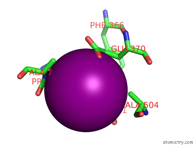
Mono view
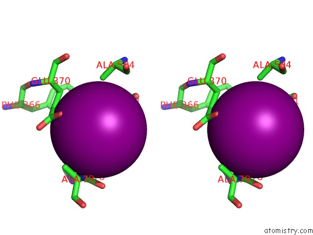
Stereo pair view
Mono view
Stereo pair view
A full contact list of Iodine with other atoms in the I binding
site number 1 of Crystal Structure of Leut Bound to L-Selenomethionine in Space Group C2 From Lipid Bicelles within 5.0Å range:
|
Reference:
H.Wang,
J.Elferich,
E.Gouaux.
Structures of Leut in Bicelles Define Conformation and Substrate Binding in A Membrane-Like Context. Nat.Struct.Mol.Biol. V. 19 212 2012.
ISSN: ISSN 1545-9993
PubMed: 22245965
DOI: 10.1038/NSMB.2215
Page generated: Fri Aug 8 15:42:33 2025
ISSN: ISSN 1545-9993
PubMed: 22245965
DOI: 10.1038/NSMB.2215
Last articles
I in 4XR3I in 4XYZ
I in 4XXS
I in 4XR2
I in 4XQD
I in 4XQ4
I in 4XR0
I in 4XQW
I in 4XPV
I in 4XNW