Iodine »
PDB 3usl-3zzz »
3vtk »
Iodine in PDB 3vtk: Thymidine Kinase From Herpes Simplex Virus Type 1 in Complex with Adp and 5-Iodo-Deoxyuridine-Monophosphate
Enzymatic activity of Thymidine Kinase From Herpes Simplex Virus Type 1 in Complex with Adp and 5-Iodo-Deoxyuridine-Monophosphate
All present enzymatic activity of Thymidine Kinase From Herpes Simplex Virus Type 1 in Complex with Adp and 5-Iodo-Deoxyuridine-Monophosphate:
2.7.1.21;
2.7.1.21;
Protein crystallography data
The structure of Thymidine Kinase From Herpes Simplex Virus Type 1 in Complex with Adp and 5-Iodo-Deoxyuridine-Monophosphate, PDB code: 3vtk
was solved by
K.Wild,
G.E.Schulz,
with X-Ray Crystallography technique. A brief refinement statistics is given in the table below:
| Resolution Low / High (Å) | 10.00 / 3.00 |
| Space group | I 41 |
| Cell size a, b, c (Å), α, β, γ (°) | 83.700, 83.700, 156.300, 90.00, 90.00, 90.00 |
| R / Rfree (%) | 22.2 / 30.5 |
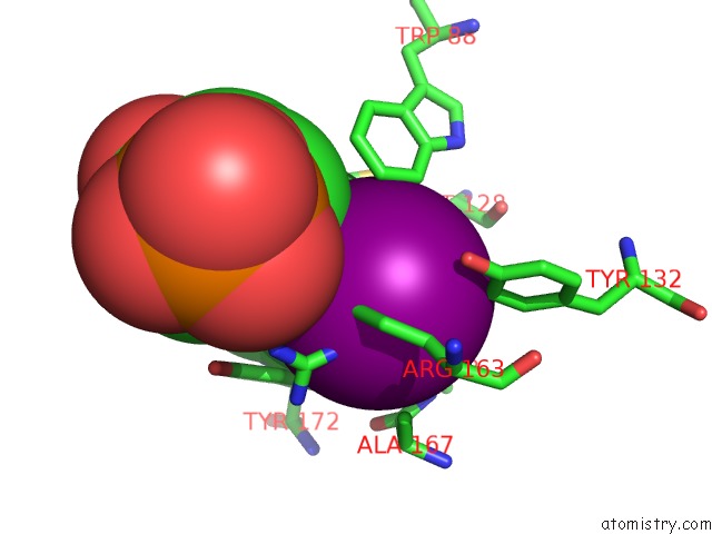
Iodine Binding Sites:
The binding sites of Iodine atom in the Thymidine Kinase From Herpes Simplex Virus Type 1 in Complex with Adp and 5-Iodo-Deoxyuridine-Monophosphate
(pdb code 3vtk). This binding sites where shown within
5.0 Angstroms radius around Iodine atom.
In total only one binding site of Iodine was determined in the Thymidine Kinase From Herpes Simplex Virus Type 1 in Complex with Adp and 5-Iodo-Deoxyuridine-Monophosphate, PDB code: 3vtk:
In total only one binding site of Iodine was determined in the Thymidine Kinase From Herpes Simplex Virus Type 1 in Complex with Adp and 5-Iodo-Deoxyuridine-Monophosphate, PDB code: 3vtk:
Iodine binding site 1 out of 1 in 3vtk
Go back to
Iodine binding site 1 out
of 1 in the Thymidine Kinase From Herpes Simplex Virus Type 1 in Complex with Adp and 5-Iodo-Deoxyuridine-Monophosphate
Mono view
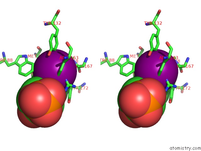
Stereo pair view
Mono view
Stereo pair view
A full contact list of Iodine with other atoms in the I binding
site number 1 of Thymidine Kinase From Herpes Simplex Virus Type 1 in Complex with Adp and 5-Iodo-Deoxyuridine-Monophosphate within 5.0Å range:
|
Reference:
K.Wild,
T.Bohner,
G.Folkers,
G.E.Schulz.
The Structures of Thymidine Kinase From Herpes Simplex Virus Type 1 in Complex with Substrates and A Substrate Analogue. Protein Sci. V. 6 2097 1997.
ISSN: ISSN 0961-8368
PubMed: 9336833
Page generated: Sun Aug 11 17:06:34 2024
ISSN: ISSN 0961-8368
PubMed: 9336833
Last articles
Zn in 9MJ5Zn in 9HNW
Zn in 9G0L
Zn in 9FNE
Zn in 9DZN
Zn in 9E0I
Zn in 9D32
Zn in 9DAK
Zn in 8ZXC
Zn in 8ZUF