Iodine »
PDB 3usl-3zzz »
3wgw »
Iodine in PDB 3wgw: Structure of Pcna Bound to A Small Molecule Inhibitor
Protein crystallography data
The structure of Structure of Pcna Bound to A Small Molecule Inhibitor, PDB code: 3wgw
was solved by
H.Hashimoto,
with X-Ray Crystallography technique. A brief refinement statistics is given in the table below:
| Resolution Low / High (Å) | 20.00 / 2.80 |
| Space group | P 41 3 2 |
| Cell size a, b, c (Å), α, β, γ (°) | 192.173, 192.173, 192.173, 90.00, 90.00, 90.00 |
| R / Rfree (%) | 18.2 / 22.1 |
Iodine Binding Sites:
The binding sites of Iodine atom in the Structure of Pcna Bound to A Small Molecule Inhibitor
(pdb code 3wgw). This binding sites where shown within
5.0 Angstroms radius around Iodine atom.
In total 6 binding sites of Iodine where determined in the Structure of Pcna Bound to A Small Molecule Inhibitor, PDB code: 3wgw:
Jump to Iodine binding site number: 1; 2; 3; 4; 5; 6;
In total 6 binding sites of Iodine where determined in the Structure of Pcna Bound to A Small Molecule Inhibitor, PDB code: 3wgw:
Jump to Iodine binding site number: 1; 2; 3; 4; 5; 6;
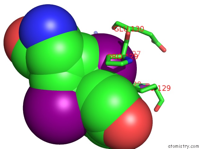
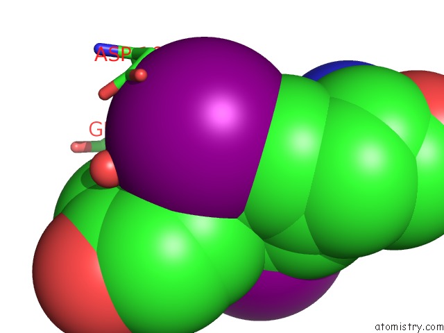
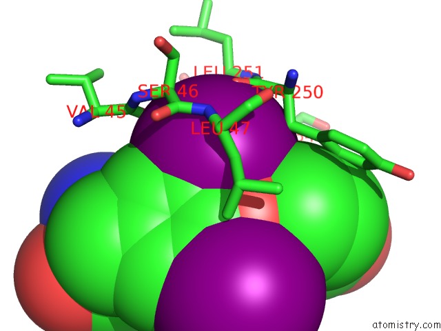
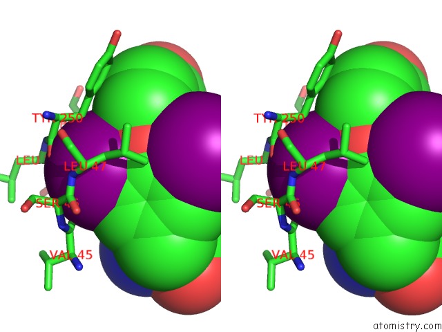
Iodine binding site 1 out of 6 in 3wgw
Go back to
Iodine binding site 1 out
of 6 in the Structure of Pcna Bound to A Small Molecule Inhibitor
Mono view
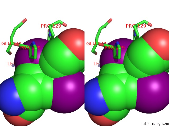
Stereo pair view
Mono view
Stereo pair view
A full contact list of Iodine with other atoms in the I binding
site number 1 of Structure of Pcna Bound to A Small Molecule Inhibitor within 5.0Å range:
|
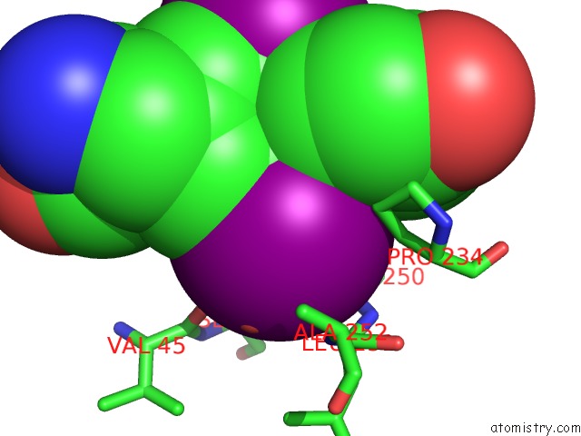
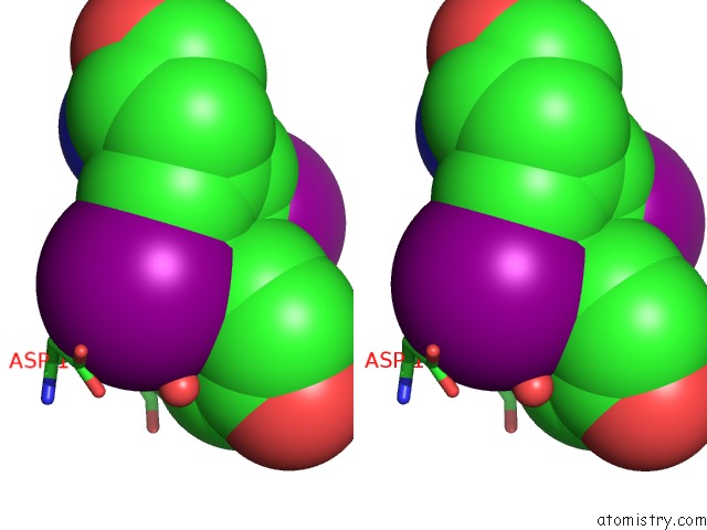
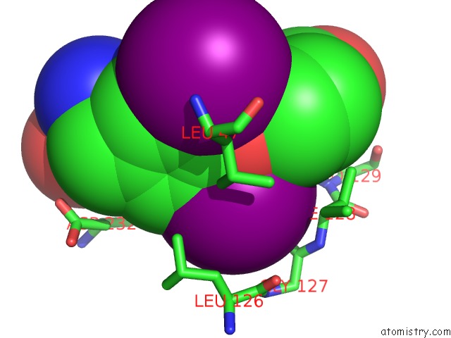
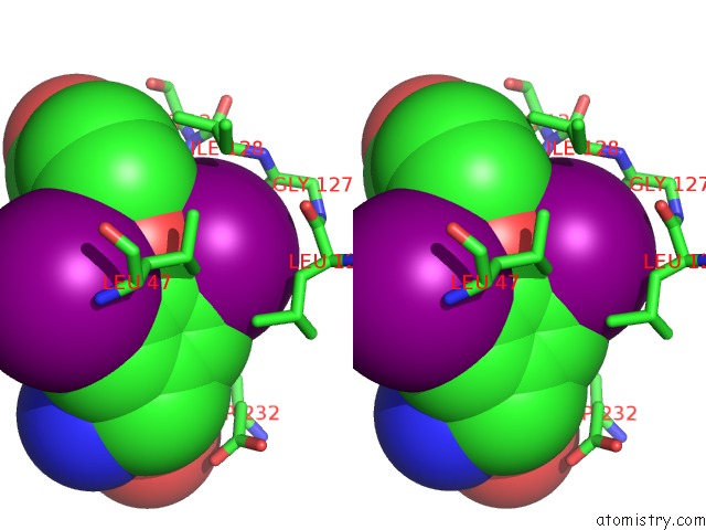
Iodine binding site 2 out of 6 in 3wgw
Go back to
Iodine binding site 2 out
of 6 in the Structure of Pcna Bound to A Small Molecule Inhibitor
Mono view
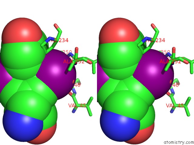
Stereo pair view
Mono view
Stereo pair view
A full contact list of Iodine with other atoms in the I binding
site number 2 of Structure of Pcna Bound to A Small Molecule Inhibitor within 5.0Å range:
|
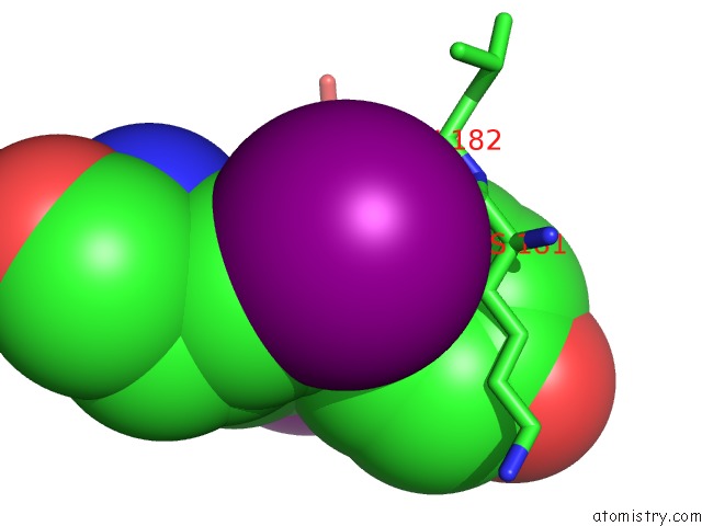
Iodine binding site 3 out of 6 in 3wgw
Go back to
Iodine binding site 3 out
of 6 in the Structure of Pcna Bound to A Small Molecule Inhibitor
Mono view
Stereo pair view
Mono view
Stereo pair view
A full contact list of Iodine with other atoms in the I binding
site number 3 of Structure of Pcna Bound to A Small Molecule Inhibitor within 5.0Å range:
|
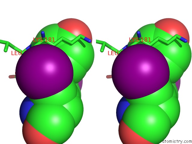
Iodine binding site 4 out of 6 in 3wgw
Go back to
Iodine binding site 4 out
of 6 in the Structure of Pcna Bound to A Small Molecule Inhibitor
Mono view
Stereo pair view
Mono view
Stereo pair view
A full contact list of Iodine with other atoms in the I binding
site number 4 of Structure of Pcna Bound to A Small Molecule Inhibitor within 5.0Å range:
|
Iodine binding site 5 out of 6 in 3wgw
Go back to
Iodine binding site 5 out
of 6 in the Structure of Pcna Bound to A Small Molecule Inhibitor
Mono view
Stereo pair view
Mono view
Stereo pair view
A full contact list of Iodine with other atoms in the I binding
site number 5 of Structure of Pcna Bound to A Small Molecule Inhibitor within 5.0Å range:
|
Iodine binding site 6 out of 6 in 3wgw
Go back to
Iodine binding site 6 out
of 6 in the Structure of Pcna Bound to A Small Molecule Inhibitor
Mono view
Stereo pair view
Mono view
Stereo pair view
A full contact list of Iodine with other atoms in the I binding
site number 6 of Structure of Pcna Bound to A Small Molecule Inhibitor within 5.0Å range:
|
Reference:
A.Inoue,
S.Kikuchi,
A.Hishiki,
Y.Shao,
R.Heath,
B.J.Evison,
M.Actis,
C.E.Canman,
H.Hashimoto,
N.Fujii.
A Small Molecule Inhibitor of Monoubiquitinated Proliferating Cell Nuclear Antigen (Pcna) Inhibits Repair of Interstrand Dna Crosslink, Enhances Dna Double-Strand Break, and Sensitizes Cancer Cells to Cisplatin J.Biol.Chem. 2014.
ISSN: ESSN 1083-351X
DOI: 10.1074/JBC.M113.520429
Page generated: Sun Aug 11 17:10:03 2024
ISSN: ESSN 1083-351X
DOI: 10.1074/JBC.M113.520429
Last articles
Zn in 9MJ5Zn in 9HNW
Zn in 9G0L
Zn in 9FNE
Zn in 9DZN
Zn in 9E0I
Zn in 9D32
Zn in 9DAK
Zn in 8ZXC
Zn in 8ZUF